Alyssa M Pereslete, Melissa E Hughes, Alyssa R Martin, Janet Files, Kyleen Nguyen, Lauren Buckley, Ashka Patel, Abigail Moore, Eric P Winer, Deborah Dillon, Tianyu Li, Sara M Tolaney, Nancy U Lin, Sarah L Sammons
{"title":"Analysis of HER2 expression changes from breast primary to brain metastases and the impact of HER2-low expression on overall survival.","authors":"Alyssa M Pereslete, Melissa E Hughes, Alyssa R Martin, Janet Files, Kyleen Nguyen, Lauren Buckley, Ashka Patel, Abigail Moore, Eric P Winer, Deborah Dillon, Tianyu Li, Sara M Tolaney, Nancy U Lin, Sarah L Sammons","doi":"10.1093/neuonc/noae163","DOIUrl":null,"url":null,"abstract":"<p><strong>Background: </strong>There are limited data regarding HER2-low expression dynamics between matched primary tumors and brain metastases (BrMs) in breast cancer. HER2-low expression has emerged as a new therapeutic biomarker for highly active antibody-drug conjugates with emerging intracranial activity.</p><p><strong>Methods: </strong>Patients with metastatic breast cancer and BrMs seen at an NCI-designated center between 2003 and 2023 were identified. HER2 expression was defined as HER2-positive (3+, 2+/ISH amplified), HER2-low (1+, 2+/ISH negative), or HER2-0 by ASCO-CAP guidelines. Estrogen receptor (ER) status was defined as ER ≥1%. Multivariate survival analyses by Cox proportional hazard models were determined from the time of BrM resection to death or last follow-up between the 3 subtypes, controlling for ER and age.</p><p><strong>Results: </strong>Among 197 matched primary and resected BrMs, 81% exhibited HER2 expression in the brain: 61% HER2-positive, 20% HER2-low, and 19% HER2-0. Concordance was high in HER2-positive primary tumors with 100% retaining HER2 expression (97% retained HER2-positive expression and 2.7% switched to HER2-low). HER2-0 primaries frequently showed HER2 gain in BrMs to HER2-low (35%) or HER2-positive (5.4%) status. Among 48 HER2-low primary tumors, 52% were discordant for HER2 status in the brain with 21% testing HER2-positive and 31% testing HER2-0. In adjusted analyses, patients with HER2-positive BrMs had significantly lower death risk than patients with HER2-low BrMs (HR = 0.41, P = .0006); no difference was observed between HER2-0 and HER2-low.</p><p><strong>Conclusions: </strong>In this retrospective analysis, HER2 expression is common in breast cancer BrMs, emphasizing the need for improved, noninvasive diagnostics. Patients with HER2-low and HER2-0 BrMs face inferior survival, presenting an unmet clinical need.</p>","PeriodicalId":19377,"journal":{"name":"Neuro-oncology","volume":" ","pages":"184-194"},"PeriodicalIF":13.4000,"publicationDate":"2025-01-12","publicationTypes":"Journal Article","fieldsOfStudy":null,"isOpenAccess":false,"openAccessPdf":"https://www.ncbi.nlm.nih.gov/pmc/articles/PMC11726339/pdf/","citationCount":"0","resultStr":null,"platform":"Semanticscholar","paperid":null,"PeriodicalName":"Neuro-oncology","FirstCategoryId":"3","ListUrlMain":"https://doi.org/10.1093/neuonc/noae163","RegionNum":1,"RegionCategory":"医学","ArticlePicture":[],"TitleCN":null,"AbstractTextCN":null,"PMCID":null,"EPubDate":"","PubModel":"","JCR":"Q1","JCRName":"CLINICAL NEUROLOGY","Score":null,"Total":0}
引用次数: 0
Abstract
Background: There are limited data regarding HER2-low expression dynamics between matched primary tumors and brain metastases (BrMs) in breast cancer. HER2-low expression has emerged as a new therapeutic biomarker for highly active antibody-drug conjugates with emerging intracranial activity.
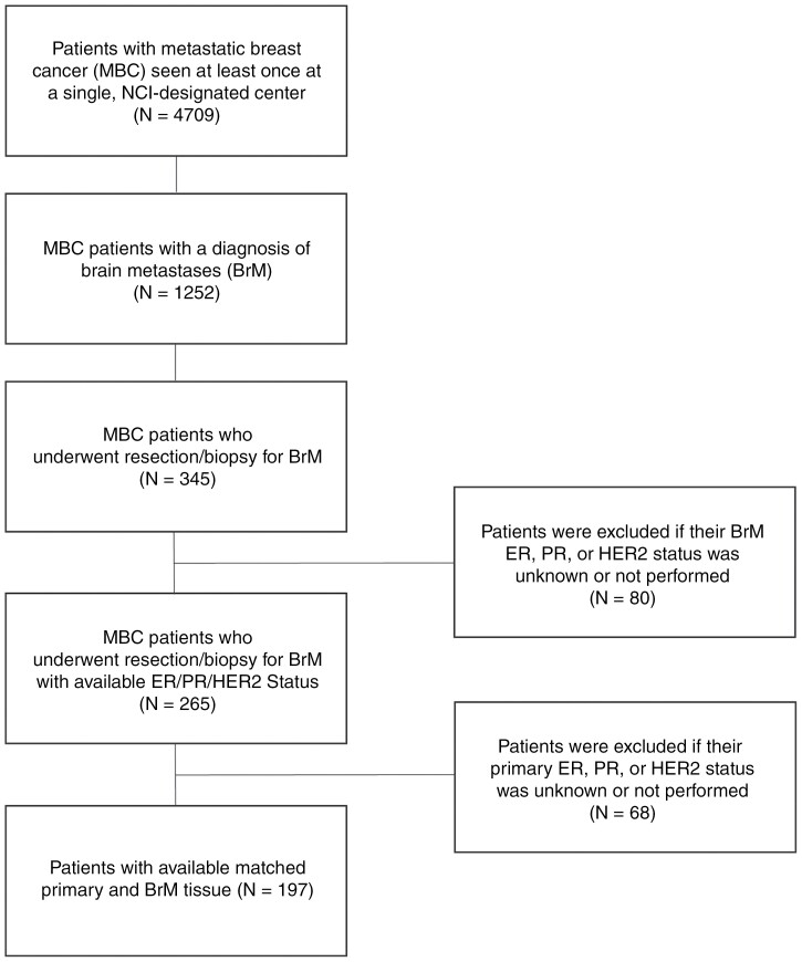
Methods: Patients with metastatic breast cancer and BrMs seen at an NCI-designated center between 2003 and 2023 were identified. HER2 expression was defined as HER2-positive (3+, 2+/ISH amplified), HER2-low (1+, 2+/ISH negative), or HER2-0 by ASCO-CAP guidelines. Estrogen receptor (ER) status was defined as ER ≥1%. Multivariate survival analyses by Cox proportional hazard models were determined from the time of BrM resection to death or last follow-up between the 3 subtypes, controlling for ER and age.
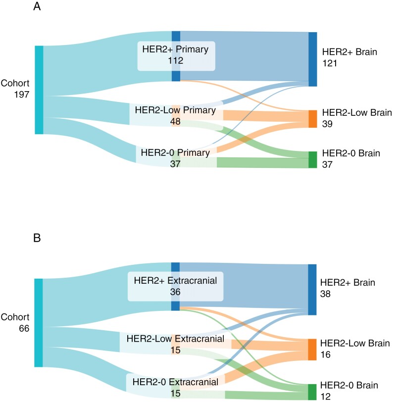
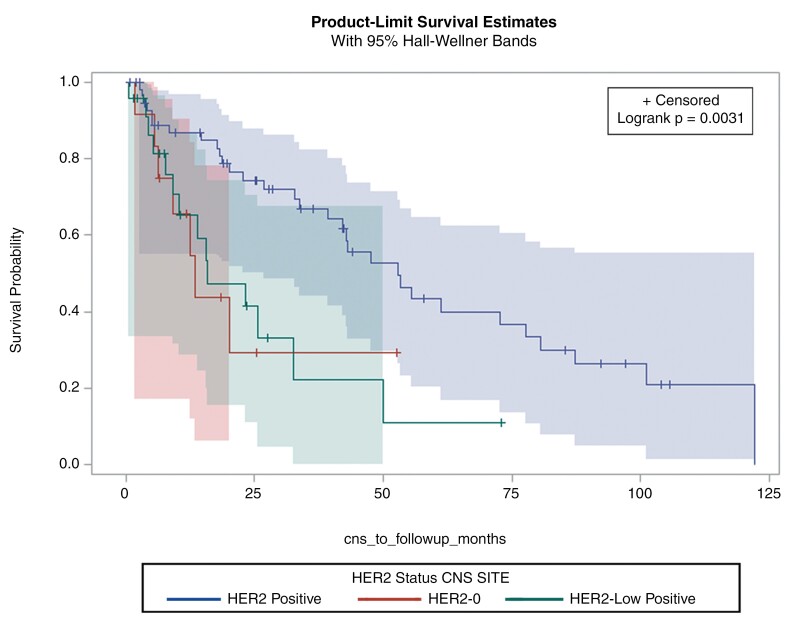
Results: Among 197 matched primary and resected BrMs, 81% exhibited HER2 expression in the brain: 61% HER2-positive, 20% HER2-low, and 19% HER2-0. Concordance was high in HER2-positive primary tumors with 100% retaining HER2 expression (97% retained HER2-positive expression and 2.7% switched to HER2-low). HER2-0 primaries frequently showed HER2 gain in BrMs to HER2-low (35%) or HER2-positive (5.4%) status. Among 48 HER2-low primary tumors, 52% were discordant for HER2 status in the brain with 21% testing HER2-positive and 31% testing HER2-0. In adjusted analyses, patients with HER2-positive BrMs had significantly lower death risk than patients with HER2-low BrMs (HR = 0.41, P = .0006); no difference was observed between HER2-0 and HER2-low.
Conclusions: In this retrospective analysis, HER2 expression is common in breast cancer BrMs, emphasizing the need for improved, noninvasive diagnostics. Patients with HER2-low and HER2-0 BrMs face inferior survival, presenting an unmet clinical need.
期刊介绍:
Neuro-Oncology, the official journal of the Society for Neuro-Oncology, has been published monthly since January 2010. Affiliated with the Japan Society for Neuro-Oncology and the European Association of Neuro-Oncology, it is a global leader in the field.
The journal is committed to swiftly disseminating high-quality information across all areas of neuro-oncology. It features peer-reviewed articles, reviews, symposia on various topics, abstracts from annual meetings, and updates from neuro-oncology societies worldwide.


 分享
分享
 求助内容:
求助内容: 应助结果提醒方式:
 扫码关注我们
