PSMD2 overexpression as a biomarker for resistance and prognosis in renal cell carcinoma treated with immune checkpoint and tyrosine kinase inhibitors.
{"title":"PSMD2 overexpression as a biomarker for resistance and prognosis in renal cell carcinoma treated with immune checkpoint and tyrosine kinase inhibitors.","authors":"Xianglai Xu, Jiahao Wang, Ying Wang, Yanjun Zhu, Jiajun Wang, Jianming Guo","doi":"10.1007/s13402-024-00977-z","DOIUrl":null,"url":null,"abstract":"<p><strong>Background: </strong>Integrated immune checkpoint inhibitors (ICIs) plus tyrosine kinase inhibitors (TKIs) are now the recommended first-line therapy to manage renal cell carcinoma (mRCC). Proteasome 26S subunit non-ATPase 2 (PSMD2) overexpression in tumors has been correlated with tumor progression. Currently, mRCC lacks an established biomarker for the combination of ICI+TKI.</p><p><strong>Methods: </strong>This study involved RNA sequencing of RCC patients from two cohorts treated with ICI+TKI (ZS-MRCC and JAVELIN-Renal-101). We utilized immunohistochemistry alongside flow cytometry, aiming at assessing immune cell infiltration and functionality in high-risk localized RCC samples. Response and progression-free survival (PFS) were evaluated relying upon RECIST criteria.</p><p><strong>Results: </strong>PSMD2 was significantly overexpressed in advanced RCC and among non-responders to ICI+TKI therapy. Overexpressed PSMD2 was correlated with poor PFS in the ZS-MRCC and JAVELIN-101 cohorts. Multivariate Cox analysis validated PSMD2 as an independent PFS predictor. PSMD2 overexpression was related to a reduction in CD8<sup>+</sup> T cells, especially GZMB<sup>+</sup> CD8<sup>+</sup> T cells, besides an increase in PD1<sup>+</sup> CD4<sup>+</sup> T cells. Additionally, tumors with high PSMD2 levels showed enhanced T cell exhaustion levels and a higher regulatory T cell presence. A Machine Learning (ML) model based on PSMD2 expression and other screened factors was subsequently developed to predict the effectiveness of ICI+TKI.</p><p><strong>Conclusions: </strong>Elevated PSMD2 expression is linked to resistance and decreased PFS in mRCC patients undergoing ICI+TKI therapy. High PSMD2 levels are also associated with impaired function and increased exhaustion of tumor-infiltrating lymphocytes. An ML model incorporating PSMD2 expression could potentially identify patients who may have a higher likelihood of benefiting from ICI+TKI.</p>","PeriodicalId":49223,"journal":{"name":"Cellular Oncology","volume":" ","pages":"1943-1956"},"PeriodicalIF":4.8000,"publicationDate":"2024-10-01","publicationTypes":"Journal Article","fieldsOfStudy":null,"isOpenAccess":false,"openAccessPdf":"","citationCount":"0","resultStr":null,"platform":"Semanticscholar","paperid":null,"PeriodicalName":"Cellular Oncology","FirstCategoryId":"3","ListUrlMain":"https://doi.org/10.1007/s13402-024-00977-z","RegionNum":2,"RegionCategory":"医学","ArticlePicture":[],"TitleCN":null,"AbstractTextCN":null,"PMCID":null,"EPubDate":"2024/9/2 0:00:00","PubModel":"Epub","JCR":"Q2","JCRName":"CELL BIOLOGY","Score":null,"Total":0}
引用次数: 0
Abstract
Background: Integrated immune checkpoint inhibitors (ICIs) plus tyrosine kinase inhibitors (TKIs) are now the recommended first-line therapy to manage renal cell carcinoma (mRCC). Proteasome 26S subunit non-ATPase 2 (PSMD2) overexpression in tumors has been correlated with tumor progression. Currently, mRCC lacks an established biomarker for the combination of ICI+TKI.
Methods: This study involved RNA sequencing of RCC patients from two cohorts treated with ICI+TKI (ZS-MRCC and JAVELIN-Renal-101). We utilized immunohistochemistry alongside flow cytometry, aiming at assessing immune cell infiltration and functionality in high-risk localized RCC samples. Response and progression-free survival (PFS) were evaluated relying upon RECIST criteria.
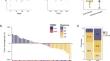
Results: PSMD2 was significantly overexpressed in advanced RCC and among non-responders to ICI+TKI therapy. Overexpressed PSMD2 was correlated with poor PFS in the ZS-MRCC and JAVELIN-101 cohorts. Multivariate Cox analysis validated PSMD2 as an independent PFS predictor. PSMD2 overexpression was related to a reduction in CD8+ T cells, especially GZMB+ CD8+ T cells, besides an increase in PD1+ CD4+ T cells. Additionally, tumors with high PSMD2 levels showed enhanced T cell exhaustion levels and a higher regulatory T cell presence. A Machine Learning (ML) model based on PSMD2 expression and other screened factors was subsequently developed to predict the effectiveness of ICI+TKI.
Conclusions: Elevated PSMD2 expression is linked to resistance and decreased PFS in mRCC patients undergoing ICI+TKI therapy. High PSMD2 levels are also associated with impaired function and increased exhaustion of tumor-infiltrating lymphocytes. An ML model incorporating PSMD2 expression could potentially identify patients who may have a higher likelihood of benefiting from ICI+TKI.
期刊介绍:
The Official Journal of the International Society for Cellular Oncology
Focuses on translational research
Addresses the conversion of cell biology to clinical applications
Cellular Oncology publishes scientific contributions from various biomedical and clinical disciplines involved in basic and translational cancer research on the cell and tissue level, technical and bioinformatics developments in this area, and clinical applications. This includes a variety of fields like genome technology, micro-arrays and other high-throughput techniques, genomic instability, SNP, DNA methylation, signaling pathways, DNA organization, (sub)microscopic imaging, proteomics, bioinformatics, functional effects of genomics, drug design and development, molecular diagnostics and targeted cancer therapies, genotype-phenotype interactions.
A major goal is to translate the latest developments in these fields from the research laboratory into routine patient management. To this end Cellular Oncology forms a platform of scientific information exchange between molecular biologists and geneticists, technical developers, pathologists, (medical) oncologists and other clinicians involved in the management of cancer patients.
In vitro studies are preferentially supported by validations in tumor tissue with clinicopathological associations.


 分享
分享
 求助内容:
求助内容: 应助结果提醒方式:
 扫码关注我们
