Emma Bertucci, Filomena Giulia Sileo, Maria Longo, Giulia Tarozzi, Martina Benuzzi, Antonio La Marca
{"title":"Isthmocele diagnosis: The optimal timing for detection","authors":"Emma Bertucci, Filomena Giulia Sileo, Maria Longo, Giulia Tarozzi, Martina Benuzzi, Antonio La Marca","doi":"10.1002/ijgo.15892","DOIUrl":null,"url":null,"abstract":"<p>The term isthmocele refers to an iatrogenic uterine defect at the site of the scar from a previous cesarean section or other isthmic tract surgery. Uterine isthmocele affects up to 70% of women with a history of previous cesarean sections, and approximately one-third of them exhibit symptoms such as menstrual spotting, abnormal uterine bleeding, or abdominal pain.<span><sup>1, 2</sup></span></p><p>Factors such as multiple cesarean sections, maternal obesity, diabetes, and retroverted uteri have been associated with the typical disruption of the myometrium at the scar.<span><sup>1</sup></span> Another potential causative factor is the closure technique of the hysterotomy, specifically, double- versus single-layer closure. A recent meta-analysis demonstrated that single-layer uterine closure had a similar incidence of isthmocele as double-layer closure 6–12 months after the cesarean section.<span><sup>3</sup></span></p><p>Accurate diagnosis is crucial as isthmocele has been linked to various gynecologic and obstetric complications such as scar pregnancy, placenta accrete spectrum disorders, and uterine rupture in subsequent pregnancies.<span><sup>4</sup></span> Additionally, women with a uterine niche may experience poorer fertility outcomes due to intrauterine fluid accumulation (mainly menstrual blood and mucus), which could lead to chronic endometrial inflammation and compromise embryo implantation.<span><sup>2</sup></span></p><p>Transvaginal ultrasonography (TVU) is considered the primary diagnostic method. Isthmocele appears as a triangular anechoic indentation communicating with the endometrial cavity within the myometrium of the lower uterine segment. The exact timing for a diagnostic ultrasound has not been yet clearly defined, with several studies conducting diagnostic procedures during the follicular phase of the menstrual cycle.<span><sup>5</sup></span> The size of the uterine niche has been shown to be different during the proliferative and secretory phases, highlighting the need to standardize the exact time of measurement.</p><p>Regarding surgical correction of the isthmocele, most authors claim that a residual myometrium ≥3 mm can be corrected by a resectoscopic approach, whereas if it is <3 mm, laparoscopic treatment is recommended.</p><p>A precise ultrasound follow-up has been carried out to refine the diagnostic timing for isthmocele.</p><p>We analyzed the diagnostic path of eight patients aged between 31 and 39, referred to our center who were subsequently diagnosed with isthmocele. Seven of our patients were diagnosed after the first cesarean section, while the remaining one was diagnosed after the second cesarean section.</p><p>All patients underwent multiple TVU examinations throughout the menstrual cycle, conducted by a single operator (EB) using Voluson E10 (General Electric, Boston, MA). Isthmocele perimeters and area were measured offline in both proliferative and secretive phases of the cycle by the same operator (EB) twice and intraobserver reliability was calculated using Pearson correlation coefficient. Ethical Committee Approval from our department was obtained in line with local law (committee: Comitato Etico Area Vasta Emilia Nord).</p><p>Comparing the uterine images of all patients through the different phases, the visualization of the isthmocele appeared clearer when the TVU was performed in the secretive phase of menstruation (Figure 1b), compared to the proliferative phase (Figure 1a). Vascularization was clearly detected in two of the total cases.</p><p>Both the mean perimeters (31 ± 5.2 vs 40.7 ± 58,2, <i>P</i> = 0.003) and the mean areas (47.9 ± 12 vs 79.8 ± 36.4, <i>P</i> = 0.010) were larger in the secretive phase (Figure 1c,d). The mean delta between the two phases was 16.9 ± 13.8 and 39.2 ± 34.4 for perimeter and area, respectively, with a mean percentage increase of 32% ± 17.4% for the perimeter and 41.1% ± 21.5% for the area.</p><p>Intraobserver reliability was high for all measurements, in particular for perimeters in proliferative (<i>r</i> = 0.8795, <i>P</i> = 0.004) and secretive (<i>r</i> = 0.9614, <i>P</i> < 0.001) phases and similarly for areas in proliferative (<i>r</i> = 0.09001, <i>P</i> = 0.002) and secretive (<i>r</i> = 0.9943, <i>P</i> < 0.001) phases.</p><p>During the secretive phase TVU revealed a more defined isthmocele in borders, echogenicity, perimeters and areas. In the second half of the menstrual cycle, the endometrium was uniformly echogenic due to mucus and glycogen in the endometrial cells. Moreover, the endometrial thickness and the enhancement of echoes allowed a better visualization of a uterine niche facilitating the isthmocele evaluation. Three patients underwent hysteroscopic treatment of uterine isthmocele and subsequently obtained spontaneous pregnancy.</p><p>Isthmocele is usually detected and characterized by two-dimensional (2D)-TVU, which is the first-line imaging approach in this diagnosis. Two recent consensus Delphi on criteria and methodology for imaging and measurements of isthmoceles in non-pregnant and early pregnancy were produced.<span><sup>6, 7</sup></span> Only one study<span><sup>8</sup></span> reported on timing of visualization of isthmocele and concluded that the best time is during the bleeding episode. However, in this study, all TVU were performed during the follicular phase of the cycle and no comparison with secretive phase was carried out. In this case series, ultrasound images were analyzed across the different stages of the menstrual cycle for visualization of the isthmocele. The measurements were reliable and the diagnosis of isthmocele was more evident during the secretive endometrial phase.</p><p>Emma Bertucci: Acquired data, designed and drafted the manuscript, critically reviewed the manuscript for important intellectual content, approved the final version to be published, and agreed to be responsible for all aspects of the work. Filomena Giulia Sileo, Maria Longo, Giulia Tarozzi, Martina Benuzzi and Antonio La Marca: Contributed to the design and draft of the manuscript. They also agree to be responsible for all aspects of the work.</p><p>None.</p><p>The authors have no conflicts of interest to declare.</p>","PeriodicalId":14164,"journal":{"name":"International Journal of Gynecology & Obstetrics","volume":"168 2","pages":"844-846"},"PeriodicalIF":2.7000,"publicationDate":"2024-09-03","publicationTypes":"Journal Article","fieldsOfStudy":null,"isOpenAccess":false,"openAccessPdf":"https://www.ncbi.nlm.nih.gov/pmc/articles/PMC11726138/pdf/","citationCount":"0","resultStr":null,"platform":"Semanticscholar","paperid":null,"PeriodicalName":"International Journal of Gynecology & Obstetrics","FirstCategoryId":"3","ListUrlMain":"https://obgyn.onlinelibrary.wiley.com/doi/10.1002/ijgo.15892","RegionNum":3,"RegionCategory":"医学","ArticlePicture":[],"TitleCN":null,"AbstractTextCN":null,"PMCID":null,"EPubDate":"","PubModel":"","JCR":"Q2","JCRName":"OBSTETRICS & GYNECOLOGY","Score":null,"Total":0}
引用次数: 0
Abstract
The term isthmocele refers to an iatrogenic uterine defect at the site of the scar from a previous cesarean section or other isthmic tract surgery. Uterine isthmocele affects up to 70% of women with a history of previous cesarean sections, and approximately one-third of them exhibit symptoms such as menstrual spotting, abnormal uterine bleeding, or abdominal pain.1, 2
Factors such as multiple cesarean sections, maternal obesity, diabetes, and retroverted uteri have been associated with the typical disruption of the myometrium at the scar.1 Another potential causative factor is the closure technique of the hysterotomy, specifically, double- versus single-layer closure. A recent meta-analysis demonstrated that single-layer uterine closure had a similar incidence of isthmocele as double-layer closure 6–12 months after the cesarean section.3
Accurate diagnosis is crucial as isthmocele has been linked to various gynecologic and obstetric complications such as scar pregnancy, placenta accrete spectrum disorders, and uterine rupture in subsequent pregnancies.4 Additionally, women with a uterine niche may experience poorer fertility outcomes due to intrauterine fluid accumulation (mainly menstrual blood and mucus), which could lead to chronic endometrial inflammation and compromise embryo implantation.2
Transvaginal ultrasonography (TVU) is considered the primary diagnostic method. Isthmocele appears as a triangular anechoic indentation communicating with the endometrial cavity within the myometrium of the lower uterine segment. The exact timing for a diagnostic ultrasound has not been yet clearly defined, with several studies conducting diagnostic procedures during the follicular phase of the menstrual cycle.5 The size of the uterine niche has been shown to be different during the proliferative and secretory phases, highlighting the need to standardize the exact time of measurement.
Regarding surgical correction of the isthmocele, most authors claim that a residual myometrium ≥3 mm can be corrected by a resectoscopic approach, whereas if it is <3 mm, laparoscopic treatment is recommended.
A precise ultrasound follow-up has been carried out to refine the diagnostic timing for isthmocele.
We analyzed the diagnostic path of eight patients aged between 31 and 39, referred to our center who were subsequently diagnosed with isthmocele. Seven of our patients were diagnosed after the first cesarean section, while the remaining one was diagnosed after the second cesarean section.
All patients underwent multiple TVU examinations throughout the menstrual cycle, conducted by a single operator (EB) using Voluson E10 (General Electric, Boston, MA). Isthmocele perimeters and area were measured offline in both proliferative and secretive phases of the cycle by the same operator (EB) twice and intraobserver reliability was calculated using Pearson correlation coefficient. Ethical Committee Approval from our department was obtained in line with local law (committee: Comitato Etico Area Vasta Emilia Nord).
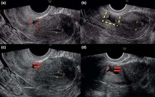
Comparing the uterine images of all patients through the different phases, the visualization of the isthmocele appeared clearer when the TVU was performed in the secretive phase of menstruation (Figure 1b), compared to the proliferative phase (Figure 1a). Vascularization was clearly detected in two of the total cases.
Both the mean perimeters (31 ± 5.2 vs 40.7 ± 58,2, P = 0.003) and the mean areas (47.9 ± 12 vs 79.8 ± 36.4, P = 0.010) were larger in the secretive phase (Figure 1c,d). The mean delta between the two phases was 16.9 ± 13.8 and 39.2 ± 34.4 for perimeter and area, respectively, with a mean percentage increase of 32% ± 17.4% for the perimeter and 41.1% ± 21.5% for the area.
Intraobserver reliability was high for all measurements, in particular for perimeters in proliferative (r = 0.8795, P = 0.004) and secretive (r = 0.9614, P < 0.001) phases and similarly for areas in proliferative (r = 0.09001, P = 0.002) and secretive (r = 0.9943, P < 0.001) phases.
During the secretive phase TVU revealed a more defined isthmocele in borders, echogenicity, perimeters and areas. In the second half of the menstrual cycle, the endometrium was uniformly echogenic due to mucus and glycogen in the endometrial cells. Moreover, the endometrial thickness and the enhancement of echoes allowed a better visualization of a uterine niche facilitating the isthmocele evaluation. Three patients underwent hysteroscopic treatment of uterine isthmocele and subsequently obtained spontaneous pregnancy.
Isthmocele is usually detected and characterized by two-dimensional (2D)-TVU, which is the first-line imaging approach in this diagnosis. Two recent consensus Delphi on criteria and methodology for imaging and measurements of isthmoceles in non-pregnant and early pregnancy were produced.6, 7 Only one study8 reported on timing of visualization of isthmocele and concluded that the best time is during the bleeding episode. However, in this study, all TVU were performed during the follicular phase of the cycle and no comparison with secretive phase was carried out. In this case series, ultrasound images were analyzed across the different stages of the menstrual cycle for visualization of the isthmocele. The measurements were reliable and the diagnosis of isthmocele was more evident during the secretive endometrial phase.
Emma Bertucci: Acquired data, designed and drafted the manuscript, critically reviewed the manuscript for important intellectual content, approved the final version to be published, and agreed to be responsible for all aspects of the work. Filomena Giulia Sileo, Maria Longo, Giulia Tarozzi, Martina Benuzzi and Antonio La Marca: Contributed to the design and draft of the manuscript. They also agree to be responsible for all aspects of the work.
None.
The authors have no conflicts of interest to declare.
峡部囊肿一词是指以前剖宫产或其他峡部手术造成的疤痕部位的医源性子宫缺损。有剖宫产史的妇女中有高达70%患有子宫峡部囊肿,其中约三分之一的妇女表现出月经斑点、子宫异常出血或腹痛等症状。1,2多次剖宫产、产妇肥胖、糖尿病和子宫内翻等因素与瘢痕处典型的子宫肌层破坏有关另一个潜在的致病因素是子宫切开术的闭合技术,特别是双层闭合与单层闭合。最近的一项荟萃分析表明,剖宫产术后6-12个月,单层子宫闭合与双层子宫闭合的峡部囊肿发生率相似。准确的诊断是至关重要的,因为峡部囊肿与各种妇科和产科并发症有关,如疤痕妊娠、胎盘增生谱系障碍和随后妊娠的子宫破裂此外,由于宫内积液(主要是经血和黏液)可能导致慢性子宫内膜炎症和胚胎着床,子宫利位妇女的生育结果可能较差。经阴道超声检查(TVU)被认为是主要的诊断方法。下段子宫肌层内,峡部肿块表现为与子宫内膜腔相通的三角形无回声凹痕。诊断超声的确切时间还没有明确规定,有几项研究在月经周期的卵泡期进行诊断子宫生态位的大小已被证明是不同的,在增殖和分泌阶段,突出需要标准化的精确测量时间。关于峡部的手术矫正,大多数作者认为,残留的肌层≥3mm可以通过切除入路进行矫正,而如果残留的肌层≥3mm,则推荐腹腔镜治疗。精确的超声随访已进行,以完善峡部膨出的诊断时机。我们分析了8例年龄在31岁至39岁之间的患者的诊断路径,这些患者随后被诊断为峡部膨出。其中7例为第一次剖宫产后确诊,1例为第二次剖宫产后确诊。所有患者在整个月经周期内均由一名操作员(EB)使用Voluson E10 (General Electric, Boston, MA)进行多次TVU检查。在周期的增殖期和隐蔽期,由同一操作员(EB)离线测量地峡周长和面积两次,并使用Pearson相关系数计算观察者内信度。伦理委员会根据当地法律获得了我们部门的批准(委员会:Comitato Etico Area Vasta Emilia Nord)。比较所有患者在不同时期的子宫图像,与增殖期(图1a)相比,在月经隐期(图1b)行TVU时,峡部的可视化更加清晰。其中2例明显发现血管化。隐匿期平均周长(31±5.2 vs 40.7±58,2,P = 0.003)和平均面积(47.9±12 vs 79.8±36.4,P = 0.010)均较大(图1c,d)。周长和面积的平均δ值分别为16.9±13.8和39.2±34.4,周长和面积的平均百分比分别增加32%±17.4%和41.1%±21.5%。所有测量结果的观察者内信度都很高,特别是在增生期(r = 0.8795, P = 0.004)和隐发期(r = 0.9614, P < 0.001)的周边,以及在增生期(r = 0.09001, P = 0.002)和隐发期(r = 0.9943, P < 0.001)的区域。在隐蔽阶段,TVU在边界、回声性、周长和面积上显示出更明确的地峡层。在月经周期的后半段,由于子宫内膜细胞中的粘液和糖原,子宫内膜回声均匀。此外,子宫内膜厚度和回声增强可以更好地显示子宫壁龛,从而促进对峡部的评估。3例患者行宫腔镜治疗子宫峡部肿大后自然妊娠。地峡囊肿通常通过二维(2D)-TVU检测和表征,这是本病诊断的一线影像学方法。最近的两个共识德尔福标准和方法成像和测量峡隔在未怀孕和妊娠早期产生。只有一项研究报道了峡部显像的时间,并得出结论,最佳时间是在出血发作期间。 然而,在本研究中,所有的TVU都是在月经周期的卵泡期进行的,没有与卵泡期进行比较。在本病例系列中,超声图像在月经周期的不同阶段进行分析,以显示峡部。测量结果是可靠的,在隐匿的子宫内膜期对峡部囊肿的诊断更为明显。Emma Bertucci:收集资料,设计并起草稿件,对稿件中重要的知识内容进行严格审查,批准最终版本出版,并同意对工作的各个方面负责。Filomena Giulia Sileo, Maria Longo, Giulia Tarozzi, Martina Benuzzi和Antonio La Marca:参与了手稿的设计和起草。作者同意对研究的所有方面负责。作者无利益冲突需要声明。
期刊介绍:
The International Journal of Gynecology & Obstetrics publishes articles on all aspects of basic and clinical research in the fields of obstetrics and gynecology and related subjects, with emphasis on matters of worldwide interest.






 京公网安备 11010802042870号
京ICP备2023020795号-1
京公网安备 11010802042870号
京ICP备2023020795号-1

 分享
分享
 求助内容:
求助内容: 应助结果提醒方式:
 扫码关注我们
