A large vertebral artery fenestration involving the distal segments associated with bilateral duplication of the superior cerebellar artery: a case report.
{"title":"A large vertebral artery fenestration involving the distal segments associated with bilateral duplication of the superior cerebellar artery: a case report.","authors":"Rabia Tasdemir, Sedat Yasin","doi":"10.1007/s00276-024-03468-w","DOIUrl":null,"url":null,"abstract":"<p><strong>Purpose: </strong>The aim of our study is to report a case of a large fenestrated vertebral artery (FVA) and bilateral duplication of the superior cerebellar artery (SCA) incidentally diagnosed using Computed Tomography Angiography (CTA) and Digital Subtraction Angiography (DSA).</p><p><strong>Case presentation: </strong>A 63-year-old female patient presenting to the neurology clinic with complaints of dizziness and balance disorder. CTA and DSA revealed a large FVA involving the V3 and V4 segments. Additionally, we observed bilateral duplicated SCAs originating from the distal basilar artery.</p><p><strong>Discussion: </strong>FVA is a rare anomaly resulting from fusion failure during the embryological period, with a reported incidence of 0.1%. FVA is often (70%) detected in the extracranial region, but it can also occur intracranially at a frequency of approximately 30%. Although various nomenclatures are used in the literature, we identified only two reports of a single fenestration encompassing the V3 and V4 segments, i.e., involving both the extracranial and intracranial regions. While duplication of the SCA is relatively common, bilateral duplication of SCA occurs at a rate of 0.9-5%.</p><p><strong>Conclusion: </strong>This case report describes an unusual case of VA fenestration involving both extracranial and intracranial segments, along with bilateral duplication of the SCAs. While rare, these findings highlight the importance of recognizing such vascular anomalies, which could be relevant for planning surgical or endovascular procedures in the posterior circulation.</p>","PeriodicalId":49461,"journal":{"name":"Surgical and Radiologic Anatomy","volume":" ","pages":"1801-1805"},"PeriodicalIF":1.2000,"publicationDate":"2024-11-01","publicationTypes":"Journal Article","fieldsOfStudy":null,"isOpenAccess":false,"openAccessPdf":"","citationCount":"0","resultStr":null,"platform":"Semanticscholar","paperid":null,"PeriodicalName":"Surgical and Radiologic Anatomy","FirstCategoryId":"3","ListUrlMain":"https://doi.org/10.1007/s00276-024-03468-w","RegionNum":4,"RegionCategory":"医学","ArticlePicture":[],"TitleCN":null,"AbstractTextCN":null,"PMCID":null,"EPubDate":"2024/9/3 0:00:00","PubModel":"Epub","JCR":"Q2","JCRName":"Medicine","Score":null,"Total":0}
引用次数: 0
Abstract
Purpose: The aim of our study is to report a case of a large fenestrated vertebral artery (FVA) and bilateral duplication of the superior cerebellar artery (SCA) incidentally diagnosed using Computed Tomography Angiography (CTA) and Digital Subtraction Angiography (DSA).
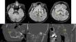
Case presentation: A 63-year-old female patient presenting to the neurology clinic with complaints of dizziness and balance disorder. CTA and DSA revealed a large FVA involving the V3 and V4 segments. Additionally, we observed bilateral duplicated SCAs originating from the distal basilar artery.
Discussion: FVA is a rare anomaly resulting from fusion failure during the embryological period, with a reported incidence of 0.1%. FVA is often (70%) detected in the extracranial region, but it can also occur intracranially at a frequency of approximately 30%. Although various nomenclatures are used in the literature, we identified only two reports of a single fenestration encompassing the V3 and V4 segments, i.e., involving both the extracranial and intracranial regions. While duplication of the SCA is relatively common, bilateral duplication of SCA occurs at a rate of 0.9-5%.
Conclusion: This case report describes an unusual case of VA fenestration involving both extracranial and intracranial segments, along with bilateral duplication of the SCAs. While rare, these findings highlight the importance of recognizing such vascular anomalies, which could be relevant for planning surgical or endovascular procedures in the posterior circulation.
期刊介绍:
Anatomy is a morphological science which cannot fail to interest the clinician. The practical application of anatomical research to clinical problems necessitates special adaptation and selectivity in choosing from numerous international works. Although there is a tendency to believe that meaningful advances in anatomy are unlikely, constant revision is necessary. Surgical and Radiologic Anatomy, the first international journal of Clinical anatomy has been created in this spirit.
Its goal is to serve clinicians, regardless of speciality-physicians, surgeons, radiologists or other specialists-as an indispensable aid with which they can improve their knowledge of anatomy. Each issue includes: Original papers, review articles, articles on the anatomical bases of medical, surgical and radiological techniques, articles of normal radiologic anatomy, brief reviews of anatomical publications of clinical interest.
Particular attention is given to high quality illustrations, which are indispensable for a better understanding of anatomical problems.
Surgical and Radiologic Anatomy is a journal written by anatomists for clinicians with a special interest in anatomy.


 分享
分享
 求助内容:
求助内容: 应助结果提醒方式:
 扫码关注我们
