{"title":"Three-dimensional computer navigation in the reconstruction of complex unilateral orbital fractures: evaluation and review of applications.","authors":"Parampreet Singh Saini, Rajesh Kumar, Manu Saini, Tarush Gupta, Sunil Gaba, Ramesh Kumar Sharma","doi":"10.7181/acfs.2024.00143","DOIUrl":null,"url":null,"abstract":"<p><strong>Background: </strong>The eyes are the central aesthetic unit of the face. Maxillofacial trauma can alter facial proportions and affect visual function with varying degrees of severity. Conventional approaches to reconstruction have numerous limitations, making the process challenging. The primary objective of this study was to evaluate the application of three-dimensional (3D) navigation in complex unilateral orbital reconstruction.</p><p><strong>Methods: </strong>A prospective cohort study was conducted over 19 months (January 2020 to July 2021), with consecutive enrollment of 12 patients who met the inclusion criteria. Each patient was followed for a minimum period of 6 months. The principal investigator carried out a comparative analysis of several factors, including fracture morphology, orbital volume, globe projection, diplopia, facial morphic changes, lid retraction, and infraorbital nerve hypoesthesia.</p><p><strong>Results: </strong>Nine patients had impure orbital fractures, while the remainder had pure fractures. The median orbital volume on the normal side (30.12 cm3; interquartile range [IQR], 28.45-30.64) was comparable to that of the reconstructed orbit (29.67 cm3; IQR, 27.92-31.52). Diplopia improved significantly (T(10) = 2.667, p = 0.02), although there was no statistically significant improvement in globe projection. Gross symmetry of facial landmarks was achieved, with comparable facial width-to-height ratio and palpebral fissure lengths. Two patients reported infraorbital hypoesthesia at presentation, which persisted at the 6-month follow-up. Additionally, five patients developed lower lid retraction (1-2 mm), and one experienced implant impingement at the infraorbital border.</p><p><strong>Conclusion: </strong>Our study provides level II evidence supporting the use of 3D navigation to improve surgical outcomes in complex orbital reconstruction.</p>","PeriodicalId":52238,"journal":{"name":"Archives of Craniofacial Surgery","volume":"25 4","pages":"161-170"},"PeriodicalIF":0.0000,"publicationDate":"2024-08-01","publicationTypes":"Journal Article","fieldsOfStudy":null,"isOpenAccess":false,"openAccessPdf":"https://www.ncbi.nlm.nih.gov/pmc/articles/PMC11374521/pdf/","citationCount":"0","resultStr":null,"platform":"Semanticscholar","paperid":null,"PeriodicalName":"Archives of Craniofacial Surgery","FirstCategoryId":"1085","ListUrlMain":"https://doi.org/10.7181/acfs.2024.00143","RegionNum":0,"RegionCategory":null,"ArticlePicture":[],"TitleCN":null,"AbstractTextCN":null,"PMCID":null,"EPubDate":"2024/8/20 0:00:00","PubModel":"Epub","JCR":"Q2","JCRName":"Medicine","Score":null,"Total":0}
引用次数: 0
Abstract
Background: The eyes are the central aesthetic unit of the face. Maxillofacial trauma can alter facial proportions and affect visual function with varying degrees of severity. Conventional approaches to reconstruction have numerous limitations, making the process challenging. The primary objective of this study was to evaluate the application of three-dimensional (3D) navigation in complex unilateral orbital reconstruction.
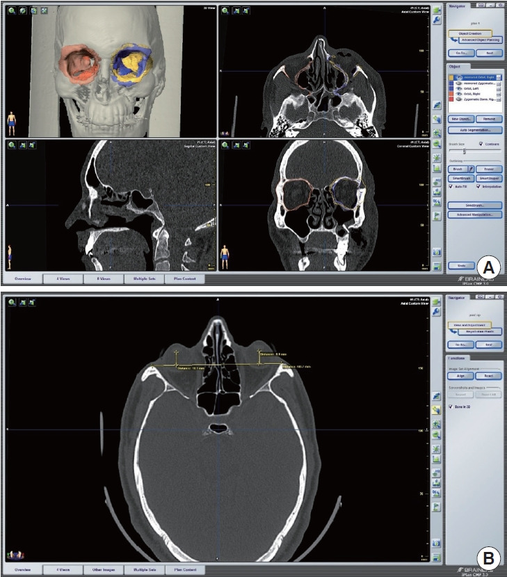
Methods: A prospective cohort study was conducted over 19 months (January 2020 to July 2021), with consecutive enrollment of 12 patients who met the inclusion criteria. Each patient was followed for a minimum period of 6 months. The principal investigator carried out a comparative analysis of several factors, including fracture morphology, orbital volume, globe projection, diplopia, facial morphic changes, lid retraction, and infraorbital nerve hypoesthesia.
Results: Nine patients had impure orbital fractures, while the remainder had pure fractures. The median orbital volume on the normal side (30.12 cm3; interquartile range [IQR], 28.45-30.64) was comparable to that of the reconstructed orbit (29.67 cm3; IQR, 27.92-31.52). Diplopia improved significantly (T(10) = 2.667, p = 0.02), although there was no statistically significant improvement in globe projection. Gross symmetry of facial landmarks was achieved, with comparable facial width-to-height ratio and palpebral fissure lengths. Two patients reported infraorbital hypoesthesia at presentation, which persisted at the 6-month follow-up. Additionally, five patients developed lower lid retraction (1-2 mm), and one experienced implant impingement at the infraorbital border.
Conclusion: Our study provides level II evidence supporting the use of 3D navigation to improve surgical outcomes in complex orbital reconstruction.


 分享
分享
 求助内容:
求助内容: 应助结果提醒方式:
 扫码关注我们
