George Triantafyllou, Nicol Zielinska, Maria Piagkou, Krzysztof Koptas, Łukasz Olewnik
{"title":"A three-headed plantaris muscle with a bipartite insertion of its two accessory heads","authors":"George Triantafyllou, Nicol Zielinska, Maria Piagkou, Krzysztof Koptas, Łukasz Olewnik","doi":"10.1007/s12565-024-00794-2","DOIUrl":null,"url":null,"abstract":"<div><p>The plantaris muscle consists of a small muscular and a long tendinous part and is located at the superficial compartment of the posterior leg. The purpose of the current cadaveric report is to describe a rare variant of the plantaris muscle. During a routine dissection, a three-headed plantaris with two accessory heads was identified with a variant insertion of the two accessory heads. All heads originated from the femur popliteal surface, independently the one from the other. The first head contributed to the long and thin calcaneal tendon, and the two accessory heads were mainly inserted via their musculoaponeurotic expansion into the medial femoral condyle. The plantaris muscle morphological variability has been extensively studied lately. The incidence of the two-headed muscle has been estimated at 1.6%, while the three-headed muscle corresponds to an even rarer variation. This is the third case reported in the English literature, while the insertion of the two accessory heads has never been described before.</p></div>","PeriodicalId":7816,"journal":{"name":"Anatomical Science International","volume":"100 1","pages":"133 - 137"},"PeriodicalIF":1.7000,"publicationDate":"2024-09-04","publicationTypes":"Journal Article","fieldsOfStudy":null,"isOpenAccess":false,"openAccessPdf":"","citationCount":"0","resultStr":null,"platform":"Semanticscholar","paperid":null,"PeriodicalName":"Anatomical Science International","FirstCategoryId":"3","ListUrlMain":"https://link.springer.com/article/10.1007/s12565-024-00794-2","RegionNum":4,"RegionCategory":"医学","ArticlePicture":[],"TitleCN":null,"AbstractTextCN":null,"PMCID":null,"EPubDate":"","PubModel":"","JCR":"Q3","JCRName":"ANATOMY & MORPHOLOGY","Score":null,"Total":0}
引用次数: 0
Abstract
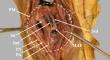
The plantaris muscle consists of a small muscular and a long tendinous part and is located at the superficial compartment of the posterior leg. The purpose of the current cadaveric report is to describe a rare variant of the plantaris muscle. During a routine dissection, a three-headed plantaris with two accessory heads was identified with a variant insertion of the two accessory heads. All heads originated from the femur popliteal surface, independently the one from the other. The first head contributed to the long and thin calcaneal tendon, and the two accessory heads were mainly inserted via their musculoaponeurotic expansion into the medial femoral condyle. The plantaris muscle morphological variability has been extensively studied lately. The incidence of the two-headed muscle has been estimated at 1.6%, while the three-headed muscle corresponds to an even rarer variation. This is the third case reported in the English literature, while the insertion of the two accessory heads has never been described before.
期刊介绍:
The official English journal of the Japanese Association of Anatomists, Anatomical Science International (formerly titled Kaibogaku Zasshi) publishes original research articles dealing with morphological sciences.
Coverage in the journal includes molecular, cellular, histological and gross anatomical studies on humans and on normal and experimental animals, as well as functional morphological, biochemical, physiological and behavioral studies if they include morphological analysis.


 分享
分享
 求助内容:
求助内容: 应助结果提醒方式:
 扫码关注我们
