Nuket Özkavruk Eliyatkın, Akif İşlek, Selim Durmaz, Fevzi Ayyıldız, Ömer Rahman
{"title":"Can adalimumab prevent from acute effects of lipopolysaccharide induced renal injury in rats?","authors":"Nuket Özkavruk Eliyatkın, Akif İşlek, Selim Durmaz, Fevzi Ayyıldız, Ömer Rahman","doi":"10.1590/acb394624","DOIUrl":null,"url":null,"abstract":"<p><strong>Purpose: </strong>Lipopolysaccharides is well-known in the acute renal injury process. It causes widespread activation of inflammatory cascades. Tumor necrosis factor (TNF)-α and interleukin (Il)-6 are essential proinflammatory cytokines that can induce the production of other cytokines in host response. Adalimumab suppresses TNF-α, IL-1β, and IL-6. We aimed to evaluate whether adalimumab would prevent the toxicity of lipopolysaccharide on the rat renal tissue.</p><p><strong>Methods: </strong>Adult female Wistar rats were divided into four groups. To the control group, only intraperitoneal saline injection procedure was carried out. For adalimumab group, adalimumab was injected at a dose for two days. For lipopolysaccharide group, animals were injected with lipopolysaccharide (a dose). For lipopolysaccharide-adalimumab group, animals were given adalimumab treatment before the injection of lipopolysaccharide. Histopathological changes and immunohistochemical analysis for TNF-α and IL-6 were determined.</p><p><strong>Results: </strong>The pathological changes and immunohistochemical staining for TNF-α or IL-6 were similar for control and adalimumab groups (p > 0.05). The lipopolysaccharide group had significantly higher distorted features in the renal tissues (p < 0.001), and also significantly prominent immunohistochemical staining for TNF-α or IL-6 (0.003), compared to the control group. No severe pathological feature was detected in the lipopolysaccharide-adalimumab group, but moderate necrosis was found in all cases (p = 0.003). TNF-α staining and IL-6 staining in the lipopolysaccharide group was found to significantly prominent compared to lipopolysaccharide-adalimumab group (p = 0.013).</p><p><strong>Conclusions: </strong>Because of its anti-inflammatory property, adalimumab pretreatment may have protective effects on experimental kidney injury. Adalimumab could be considered as a protective agent to acute effects of lipopolysaccharide induced renal injury.</p>","PeriodicalId":93850,"journal":{"name":"Acta cirurgica brasileira","volume":"39 ","pages":"e394624"},"PeriodicalIF":1.3000,"publicationDate":"2024-09-02","publicationTypes":"Journal Article","fieldsOfStudy":null,"isOpenAccess":false,"openAccessPdf":"https://www.ncbi.nlm.nih.gov/pmc/articles/PMC11368208/pdf/","citationCount":"0","resultStr":null,"platform":"Semanticscholar","paperid":null,"PeriodicalName":"Acta cirurgica brasileira","FirstCategoryId":"1085","ListUrlMain":"https://doi.org/10.1590/acb394624","RegionNum":0,"RegionCategory":null,"ArticlePicture":[],"TitleCN":null,"AbstractTextCN":null,"PMCID":null,"EPubDate":"2024/1/1 0:00:00","PubModel":"eCollection","JCR":"","JCRName":"","Score":null,"Total":0}
引用次数: 0
Abstract
Purpose: Lipopolysaccharides is well-known in the acute renal injury process. It causes widespread activation of inflammatory cascades. Tumor necrosis factor (TNF)-α and interleukin (Il)-6 are essential proinflammatory cytokines that can induce the production of other cytokines in host response. Adalimumab suppresses TNF-α, IL-1β, and IL-6. We aimed to evaluate whether adalimumab would prevent the toxicity of lipopolysaccharide on the rat renal tissue.
Methods: Adult female Wistar rats were divided into four groups. To the control group, only intraperitoneal saline injection procedure was carried out. For adalimumab group, adalimumab was injected at a dose for two days. For lipopolysaccharide group, animals were injected with lipopolysaccharide (a dose). For lipopolysaccharide-adalimumab group, animals were given adalimumab treatment before the injection of lipopolysaccharide. Histopathological changes and immunohistochemical analysis for TNF-α and IL-6 were determined.
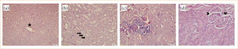
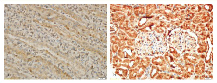
Results: The pathological changes and immunohistochemical staining for TNF-α or IL-6 were similar for control and adalimumab groups (p > 0.05). The lipopolysaccharide group had significantly higher distorted features in the renal tissues (p < 0.001), and also significantly prominent immunohistochemical staining for TNF-α or IL-6 (0.003), compared to the control group. No severe pathological feature was detected in the lipopolysaccharide-adalimumab group, but moderate necrosis was found in all cases (p = 0.003). TNF-α staining and IL-6 staining in the lipopolysaccharide group was found to significantly prominent compared to lipopolysaccharide-adalimumab group (p = 0.013).
Conclusions: Because of its anti-inflammatory property, adalimumab pretreatment may have protective effects on experimental kidney injury. Adalimumab could be considered as a protective agent to acute effects of lipopolysaccharide induced renal injury.


 分享
分享

 求助内容:
求助内容: 应助结果提醒方式:
 扫码关注我们
