{"title":"Activation of the LKB1/AMPK/HIF-1α Pathway by Metformin to Promote Neovascularisation in Cerebral Ischaemia","authors":"Hongguang Chen, Yuting Yuan, Yue Zhang, Xiufen Liu, Qingjie Chen, Chao Liu, Qing Yao","doi":"10.1007/s11064-024-04235-4","DOIUrl":null,"url":null,"abstract":"<div><p>As a difficult-to-treat neurological condition, cerebral ischemia is currently limited to treatments such as intravenous recombinant tissue plasminogen activator thrombolysis and thrombectomy. Metformin, a potent antidiabetic drug, has been reported to have an independent function in enhancing the prognosis of stroke patients, in addition to its glucose-lowering effects. However, the mechanism of action of metformin in this context remains unclear. In vivo, a rat model of permanent middle cerebral artery occlusion was established, and after administration of a low dose of 10.5 mg/mL metformin, infarct area was measured by TTC staining, and cortical blood flow was determined by laser Doppler imaging. In vitro, the study established human umbilical vein endothelial cells treated with cobalt chloride. Immunofluorescence, immunohistochemistry, and Western blot experiments were performed to observe the expression of angiogenic factors, tight junction proteins, and apoptotic factors. A TUNEL assay was utilized to appraise cell death by apoptosis. A tube formation assay and scratch assay were conducted to determine the endothelial neovascularization status. Animal experiments have revealed that the administration of the AMPK activator metformin significantly reduced the infarct area, promoted the expression of angiogenic factors, and maintained the stability of tight junction proteins in endothelial cells. Moreover, metformin reduces nerve cells apoptosis by affecting the expression of the apoptotic protein cleaved-caspase3 via the HIF-1α pathway. In vitro, the LKB1/AMPK signaling pathway is activated after hypoxic stimulation, attaining its peak within the early stages of hypoxia (1–12 h) and gradually weakening thereafter. The administration of AMPK pharmacological agonists (between 36 and 48 h) can enhance AMPK activity, which can lead to the expression of angiogenic factors, maintain the stability of tight-junction proteins in endothelial cells, and facilitate endothelial cell migration and vascular structure formation. Conversely, the AMPK inhibitors exert the opposite effects. The activation of the LKB1/AMPK/HIF-1α signaling pathway by metformin in cerebral ischemia contributes to angiogenesis, promotes tissue repair in the injured area, and enhances neurologically functional symptoms.</p></div>","PeriodicalId":719,"journal":{"name":"Neurochemical Research","volume":"49 12","pages":"3263 - 3276"},"PeriodicalIF":3.8000,"publicationDate":"2024-09-06","publicationTypes":"Journal Article","fieldsOfStudy":null,"isOpenAccess":false,"openAccessPdf":"","citationCount":"0","resultStr":null,"platform":"Semanticscholar","paperid":null,"PeriodicalName":"Neurochemical Research","FirstCategoryId":"3","ListUrlMain":"https://link.springer.com/article/10.1007/s11064-024-04235-4","RegionNum":3,"RegionCategory":"医学","ArticlePicture":[],"TitleCN":null,"AbstractTextCN":null,"PMCID":null,"EPubDate":"","PubModel":"","JCR":"Q2","JCRName":"BIOCHEMISTRY & MOLECULAR BIOLOGY","Score":null,"Total":0}
引用次数: 0
Abstract
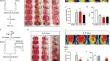
As a difficult-to-treat neurological condition, cerebral ischemia is currently limited to treatments such as intravenous recombinant tissue plasminogen activator thrombolysis and thrombectomy. Metformin, a potent antidiabetic drug, has been reported to have an independent function in enhancing the prognosis of stroke patients, in addition to its glucose-lowering effects. However, the mechanism of action of metformin in this context remains unclear. In vivo, a rat model of permanent middle cerebral artery occlusion was established, and after administration of a low dose of 10.5 mg/mL metformin, infarct area was measured by TTC staining, and cortical blood flow was determined by laser Doppler imaging. In vitro, the study established human umbilical vein endothelial cells treated with cobalt chloride. Immunofluorescence, immunohistochemistry, and Western blot experiments were performed to observe the expression of angiogenic factors, tight junction proteins, and apoptotic factors. A TUNEL assay was utilized to appraise cell death by apoptosis. A tube formation assay and scratch assay were conducted to determine the endothelial neovascularization status. Animal experiments have revealed that the administration of the AMPK activator metformin significantly reduced the infarct area, promoted the expression of angiogenic factors, and maintained the stability of tight junction proteins in endothelial cells. Moreover, metformin reduces nerve cells apoptosis by affecting the expression of the apoptotic protein cleaved-caspase3 via the HIF-1α pathway. In vitro, the LKB1/AMPK signaling pathway is activated after hypoxic stimulation, attaining its peak within the early stages of hypoxia (1–12 h) and gradually weakening thereafter. The administration of AMPK pharmacological agonists (between 36 and 48 h) can enhance AMPK activity, which can lead to the expression of angiogenic factors, maintain the stability of tight-junction proteins in endothelial cells, and facilitate endothelial cell migration and vascular structure formation. Conversely, the AMPK inhibitors exert the opposite effects. The activation of the LKB1/AMPK/HIF-1α signaling pathway by metformin in cerebral ischemia contributes to angiogenesis, promotes tissue repair in the injured area, and enhances neurologically functional symptoms.
期刊介绍:
Neurochemical Research is devoted to the rapid publication of studies that use neurochemical methodology in research on nervous system structure and function. The journal publishes original reports of experimental and clinical research results, perceptive reviews of significant problem areas in the neurosciences, brief comments of a methodological or interpretive nature, and research summaries conducted by leading scientists whose works are not readily available in English.


 分享
分享
 求助内容:
求助内容: 应助结果提醒方式:
 扫码关注我们
