Dimitrios Roukas, Evangelos Tsiambas, Despoina Spyropoulou, Maria Adamopoulou, George Tsouvelas, Sofianiki Mastronikoli, Antonella-Effrosyni Monastirioti, Anastasios Kouzoupis, Andreas Lazaris, Nikolaos Kavantzas
{"title":"Caspase 3 Expression Profiles in Meningioma Subtypes Based on Tissue Microarray Analysis.","authors":"Dimitrios Roukas, Evangelos Tsiambas, Despoina Spyropoulou, Maria Adamopoulou, George Tsouvelas, Sofianiki Mastronikoli, Antonella-Effrosyni Monastirioti, Anastasios Kouzoupis, Andreas Lazaris, Nikolaos Kavantzas","doi":"10.21873/cdp.10367","DOIUrl":null,"url":null,"abstract":"<p><strong>Background/aim: </strong>Concerning primary central nervous system neoplasms, meningiomas demonstrate the most common type in adults worldwide. Deregulation of apoptotic pathways in malignancies, including meningiomas, is correlated with chemoresistance and poor prognosis. Caspases represent crucial proteins that induce cell apoptosis. This study aimed to correlate caspase 3 protein expression levels to meningioma clinic-pathological features.</p><p><strong>Materials and methods: </strong>A set of fifty (n=50) meningioma lesions was included in the current analysis including a broad spectrum of histopathological subtypes (meningotheliomatous, psammomatus, transitional, fibrous, angiomatous, microcystic, atypical and anaplastic). Immunohistochemistry was implemented on tissue microarray cores of selected paraffin blocks by applying an anti-caspase 3 antibody. Additionally, an image analysis protocol was also performed in the corresponding immunostained slides.</p><p><strong>Results: </strong>Caspase 3 protein over-expression was detected in 17/50 (34%) cases, whereas the remaining 33 cases (66%) were characterized by medium to low levels of the molecule. Caspase 3 expression was statistically significantly associated with the grade of the analyzed tumors and the mitotic index (p=0.002, p=0.001, respectively). Caspase 3 expression status was also correlated with the histotype of the selected meningiomas (p=0.016).</p><p><strong>Conclusion: </strong>Caspase 3 demonstrated low expression levels in a significant subset of the examined meningiomas correlated with differentiation grade, mitotic activity, and partially with specific histotypes. Agents that could enhance caspase 3 expression - inducing its apoptotic activity - represent a very promising area in oncology for developing novel treatment regimens.</p>","PeriodicalId":72510,"journal":{"name":"Cancer diagnosis & prognosis","volume":"4 5","pages":"586-591"},"PeriodicalIF":0.0000,"publicationDate":"2024-09-01","publicationTypes":"Journal Article","fieldsOfStudy":null,"isOpenAccess":false,"openAccessPdf":"https://www.ncbi.nlm.nih.gov/pmc/articles/PMC11372700/pdf/","citationCount":"0","resultStr":null,"platform":"Semanticscholar","paperid":null,"PeriodicalName":"Cancer diagnosis & prognosis","FirstCategoryId":"1085","ListUrlMain":"https://doi.org/10.21873/cdp.10367","RegionNum":0,"RegionCategory":null,"ArticlePicture":[],"TitleCN":null,"AbstractTextCN":null,"PMCID":null,"EPubDate":"","PubModel":"","JCR":"","JCRName":"","Score":null,"Total":0}
引用次数: 0
Abstract
Background/aim: Concerning primary central nervous system neoplasms, meningiomas demonstrate the most common type in adults worldwide. Deregulation of apoptotic pathways in malignancies, including meningiomas, is correlated with chemoresistance and poor prognosis. Caspases represent crucial proteins that induce cell apoptosis. This study aimed to correlate caspase 3 protein expression levels to meningioma clinic-pathological features.
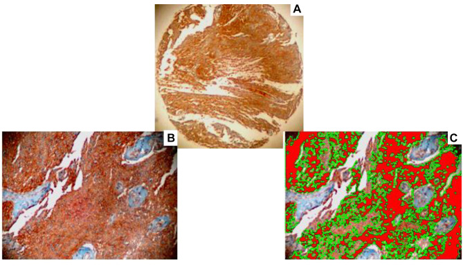
Materials and methods: A set of fifty (n=50) meningioma lesions was included in the current analysis including a broad spectrum of histopathological subtypes (meningotheliomatous, psammomatus, transitional, fibrous, angiomatous, microcystic, atypical and anaplastic). Immunohistochemistry was implemented on tissue microarray cores of selected paraffin blocks by applying an anti-caspase 3 antibody. Additionally, an image analysis protocol was also performed in the corresponding immunostained slides.
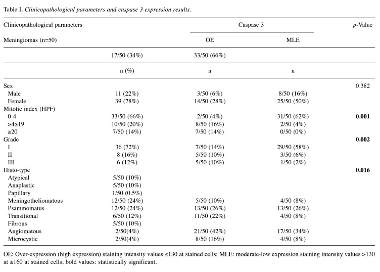
Results: Caspase 3 protein over-expression was detected in 17/50 (34%) cases, whereas the remaining 33 cases (66%) were characterized by medium to low levels of the molecule. Caspase 3 expression was statistically significantly associated with the grade of the analyzed tumors and the mitotic index (p=0.002, p=0.001, respectively). Caspase 3 expression status was also correlated with the histotype of the selected meningiomas (p=0.016).
Conclusion: Caspase 3 demonstrated low expression levels in a significant subset of the examined meningiomas correlated with differentiation grade, mitotic activity, and partially with specific histotypes. Agents that could enhance caspase 3 expression - inducing its apoptotic activity - represent a very promising area in oncology for developing novel treatment regimens.


 分享
分享

 求助内容:
求助内容: 应助结果提醒方式:
 扫码关注我们
