Andrew L. Wang , Orin Mishkit , Heather Mao , Lakshmi Arivazhagan , Tony Dong , Frances Lee , Aparajita Bhattacharya , P. Douglas Renfrew , Ann Marie Schmidt , Youssef Z. Wadghiri , Edward A. Fisher , Jin Kim Montclare
{"title":"Collagen-targeted protein nanomicelles for the imaging of non-alcoholic steatohepatitis","authors":"Andrew L. Wang , Orin Mishkit , Heather Mao , Lakshmi Arivazhagan , Tony Dong , Frances Lee , Aparajita Bhattacharya , P. Douglas Renfrew , Ann Marie Schmidt , Youssef Z. Wadghiri , Edward A. Fisher , Jin Kim Montclare","doi":"10.1016/j.actbio.2024.08.052","DOIUrl":null,"url":null,"abstract":"<div><div><em>In vivo</em> molecular imaging tools hold immense potential to drive transformative breakthroughs by enabling researchers to visualize cellular and molecular interactions in real-time and/or at high resolution. These advancements will facilitate a deeper understanding of fundamental biological processes and their dysregulation in disease states. Here, we develop and characterize a self-assembling protein nanomicelle called collagen type I binding – thermoresponsive assembled protein (Col1-TRAP) that binds tightly to type I collagen <em>in vitro</em> with nanomolar affinity. For <em>ex vivo</em> visualization, Col1-TRAP is labeled with a near-infrared fluorescent dye (NIR-Col1-TRAP). Both Col1-TRAP and NIR-Col1-TRAP display approximately a 3.8-fold greater binding to type I collagen compared to TRAP when measured by surface plasmon resonance (SPR). We present a proof-of-concept study using NIR-Col1-TRAP to detect fibrotic type I collagen deposition <em>ex vivo</em> in the livers of mice with non-alcoholic steatohepatitis (NASH). We show that NIR-Col1-TRAP demonstrates significantly decreased plasma recirculation time as well as increased liver accumulation in the NASH mice compared to mice without disease over 4 hours. As a result, NIR-Col1-TRAP shows potential as an imaging probe for NASH with <em>in vivo</em> targeting performance after injection in mice.</div></div><div><h3>Statement of significance</h3><div>Direct molecular imaging of fibrosis in NASH patients enables the diagnosis and monitoring of disease progression with greater specificity and resolution than do elastography-based methods or blood tests. In addition, protein-based imaging probes are more advantageous than alternatives due to their biodegradability and scalable biosynthesis. With the aid of computational modeling, we have designed a self-assembled protein micelle that binds to fibrillar and monomeric collagen <em>in vitro.</em> After the protein was labeled with near-infrared fluorescent dye, we injected the compound into mice fed on a NASH diet. NIR-Col1-TRAP clears from the serum faster in these mice compared to control mice, and accumulates significantly more in fibrotic livers.This work advances the development of targeted protein probes for <em>in vivo</em> fibrosis imaging.</div></div>","PeriodicalId":237,"journal":{"name":"Acta Biomaterialia","volume":"187 ","pages":"Pages 291-303"},"PeriodicalIF":9.6000,"publicationDate":"2024-10-01","publicationTypes":"Journal Article","fieldsOfStudy":null,"isOpenAccess":false,"openAccessPdf":"","citationCount":"0","resultStr":null,"platform":"Semanticscholar","paperid":null,"PeriodicalName":"Acta Biomaterialia","FirstCategoryId":"5","ListUrlMain":"https://www.sciencedirect.com/science/article/pii/S174270612400504X","RegionNum":1,"RegionCategory":"医学","ArticlePicture":[],"TitleCN":null,"AbstractTextCN":null,"PMCID":null,"EPubDate":"","PubModel":"","JCR":"Q1","JCRName":"ENGINEERING, BIOMEDICAL","Score":null,"Total":0}
引用次数: 0
Abstract
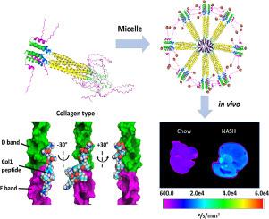
In vivo molecular imaging tools hold immense potential to drive transformative breakthroughs by enabling researchers to visualize cellular and molecular interactions in real-time and/or at high resolution. These advancements will facilitate a deeper understanding of fundamental biological processes and their dysregulation in disease states. Here, we develop and characterize a self-assembling protein nanomicelle called collagen type I binding – thermoresponsive assembled protein (Col1-TRAP) that binds tightly to type I collagen in vitro with nanomolar affinity. For ex vivo visualization, Col1-TRAP is labeled with a near-infrared fluorescent dye (NIR-Col1-TRAP). Both Col1-TRAP and NIR-Col1-TRAP display approximately a 3.8-fold greater binding to type I collagen compared to TRAP when measured by surface plasmon resonance (SPR). We present a proof-of-concept study using NIR-Col1-TRAP to detect fibrotic type I collagen deposition ex vivo in the livers of mice with non-alcoholic steatohepatitis (NASH). We show that NIR-Col1-TRAP demonstrates significantly decreased plasma recirculation time as well as increased liver accumulation in the NASH mice compared to mice without disease over 4 hours. As a result, NIR-Col1-TRAP shows potential as an imaging probe for NASH with in vivo targeting performance after injection in mice.
Statement of significance
Direct molecular imaging of fibrosis in NASH patients enables the diagnosis and monitoring of disease progression with greater specificity and resolution than do elastography-based methods or blood tests. In addition, protein-based imaging probes are more advantageous than alternatives due to their biodegradability and scalable biosynthesis. With the aid of computational modeling, we have designed a self-assembled protein micelle that binds to fibrillar and monomeric collagen in vitro. After the protein was labeled with near-infrared fluorescent dye, we injected the compound into mice fed on a NASH diet. NIR-Col1-TRAP clears from the serum faster in these mice compared to control mice, and accumulates significantly more in fibrotic livers.This work advances the development of targeted protein probes for in vivo fibrosis imaging.
期刊介绍:
Acta Biomaterialia is a monthly peer-reviewed scientific journal published by Elsevier. The journal was established in January 2005. The editor-in-chief is W.R. Wagner (University of Pittsburgh). The journal covers research in biomaterials science, including the interrelationship of biomaterial structure and function from macroscale to nanoscale. Topical coverage includes biomedical and biocompatible materials.


 分享
分享
 求助内容:
求助内容: 应助结果提醒方式:
 扫码关注我们
