Histopathologic Differential Diagnosis and Estrogen Receptor/Progesterone Receptor Immunohistochemical Evaluation of Breast Carcinoma Using a Deep Learning–Based Artificial Intelligence Architecture
Zhi Han , Shihong Ding , Baichen Liu , Yandong Tang , Xueshan Qiu , Enhua Wang , Huanyu Zhao
{"title":"Histopathologic Differential Diagnosis and Estrogen Receptor/Progesterone Receptor Immunohistochemical Evaluation of Breast Carcinoma Using a Deep Learning–Based Artificial Intelligence Architecture","authors":"Zhi Han , Shihong Ding , Baichen Liu , Yandong Tang , Xueshan Qiu , Enhua Wang , Huanyu Zhao","doi":"10.1016/j.ajpath.2024.08.011","DOIUrl":null,"url":null,"abstract":"<div><div>In breast carcinoma, invasive ductal carcinoma (IDC) is the most common histopathologic subtype, and ductal carcinoma <em>in situ</em> (DCIS) is a precursor of IDC. These two often occur concomitantly. The immunohistochemical staining of estrogen receptor (ER)/progesterone receptor (PR) in IDC/DCIS on histopathologic whole slide images (WSIs) can predict the prognosis of patients. Artificial intelligence (AI) technology has the potential to substantially reduce the interobserver variability among pathologists reading WSIs. Herein, IDC/DCIS detection was conducted by a deep learning approach, including faster region-based convolutional neural network (Faster R-CNN), RetinaNet, single-shot multibox detector 300 (SSD300), you only look once (YOLO) v3, YOLOv5, YOLOv7, YOLOv8, and Swin transformer. Their performance was estimated by mean average precision (mAP) values. Cell recognition and counting were performed using AI technology to evaluate the intensity and proportion of ER/PR-immunostained cancer cells in IDC/DCIS. A three-round ring study (RS) was conducted to assess WSIs. A database for modelling the underlying probability distribution of a data set with labels was established. YOLOv8 exhibited the highest detection performance with an mAP at 0.5 of 0.944 and an mAP at 0.5 to 0.95 of 0.790. With the assistance of YOLOv8, the scoring concordance across all pathologists was boosted to excellent in RS3 (0.970) from moderate in RS1 (0.724) and good in RS2 (0.812). Deep learning detection can be applied in the clinicopathologic field. Herein, a novel AI architecture and well-organized data set were developed to facilitate the histopathologic diagnosis of IDC/DCIS and immunostaining scoring of ER/PR.</div></div>","PeriodicalId":7623,"journal":{"name":"American Journal of Pathology","volume":"194 12","pages":"Pages 2313-2325"},"PeriodicalIF":4.9000,"publicationDate":"2024-12-01","publicationTypes":"Journal Article","fieldsOfStudy":null,"isOpenAccess":false,"openAccessPdf":"","citationCount":"0","resultStr":null,"platform":"Semanticscholar","paperid":null,"PeriodicalName":"American Journal of Pathology","FirstCategoryId":"3","ListUrlMain":"https://www.sciencedirect.com/science/article/pii/S0002944024003304","RegionNum":2,"RegionCategory":"医学","ArticlePicture":[],"TitleCN":null,"AbstractTextCN":null,"PMCID":null,"EPubDate":"2024/9/4 0:00:00","PubModel":"Epub","JCR":"Q1","JCRName":"PATHOLOGY","Score":null,"Total":0}
引用次数: 0
Abstract
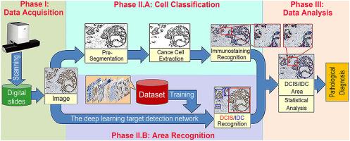
In breast carcinoma, invasive ductal carcinoma (IDC) is the most common histopathologic subtype, and ductal carcinoma in situ (DCIS) is a precursor of IDC. These two often occur concomitantly. The immunohistochemical staining of estrogen receptor (ER)/progesterone receptor (PR) in IDC/DCIS on histopathologic whole slide images (WSIs) can predict the prognosis of patients. Artificial intelligence (AI) technology has the potential to substantially reduce the interobserver variability among pathologists reading WSIs. Herein, IDC/DCIS detection was conducted by a deep learning approach, including faster region-based convolutional neural network (Faster R-CNN), RetinaNet, single-shot multibox detector 300 (SSD300), you only look once (YOLO) v3, YOLOv5, YOLOv7, YOLOv8, and Swin transformer. Their performance was estimated by mean average precision (mAP) values. Cell recognition and counting were performed using AI technology to evaluate the intensity and proportion of ER/PR-immunostained cancer cells in IDC/DCIS. A three-round ring study (RS) was conducted to assess WSIs. A database for modelling the underlying probability distribution of a data set with labels was established. YOLOv8 exhibited the highest detection performance with an mAP at 0.5 of 0.944 and an mAP at 0.5 to 0.95 of 0.790. With the assistance of YOLOv8, the scoring concordance across all pathologists was boosted to excellent in RS3 (0.970) from moderate in RS1 (0.724) and good in RS2 (0.812). Deep learning detection can be applied in the clinicopathologic field. Herein, a novel AI architecture and well-organized data set were developed to facilitate the histopathologic diagnosis of IDC/DCIS and immunostaining scoring of ER/PR.
期刊介绍:
The American Journal of Pathology, official journal of the American Society for Investigative Pathology, published by Elsevier, Inc., seeks high-quality original research reports, reviews, and commentaries related to the molecular and cellular basis of disease. The editors will consider basic, translational, and clinical investigations that directly address mechanisms of pathogenesis or provide a foundation for future mechanistic inquiries. Examples of such foundational investigations include data mining, identification of biomarkers, molecular pathology, and discovery research. Foundational studies that incorporate deep learning and artificial intelligence are also welcome. High priority is given to studies of human disease and relevant experimental models using molecular, cellular, and organismal approaches.


 分享
分享
 求助内容:
求助内容: 应助结果提醒方式:
 扫码关注我们
