Esther Collado, Adam Bardoczi, Alan B Lumsden, Zsolt Garami
{"title":"Internal Carotid Artery Hypoplasia Misidentified as Internal Carotid Artery Dissection.","authors":"Esther Collado, Adam Bardoczi, Alan B Lumsden, Zsolt Garami","doi":"10.14797/mdcvj.1417","DOIUrl":null,"url":null,"abstract":"<p><p>Agenesis or hypoplasia of the internal carotid artery (ICA) may easily be confused with dissection or occlusion. We report a case of a 24-year-old female with complaint of acute left-hand hypoesthesia and a history of occasional intermittent numbness of her right hand with myoclonic jerking. Because previous imaging studies over 2 years were interpreted as occlusion of the left ICA secondary to carotid dissection, the treating physician had prescribed anticoagulant therapy. During transcranial Doppler (TCD) examination, the spectral waveform was unexpectedly normal, prompting a repeat review of all imaging due to the TCD results. Magnetic resonance angiography (MRA) revealed the same \"flame-like\" appearance of the ICA origin. Late-phase digital subtraction angiography showed a small caliber cervical ICA (occluded at the skull base). Computed tomography demonstrated absence of the carotid canal, confirming an absent intracranial portion of the ICA and establishing a correct diagnosis of left internal carotid hypoplasia. Vascular ultrasound and TCD examinations are noninvasive and inexpensive tools that can improve the interpretation and understanding of the clinical significance of other \"static\" radiographic tests (MRA, digital subtraction angiography ). An accurate diagnosis is essential to avoid risky, aggressive treatment, such as anticoagulation for an \"absent\" dissection.</p>","PeriodicalId":39207,"journal":{"name":"Methodist DeBakey cardiovascular journal","volume":"20 1","pages":"87-93"},"PeriodicalIF":0.0000,"publicationDate":"2024-09-02","publicationTypes":"Journal Article","fieldsOfStudy":null,"isOpenAccess":false,"openAccessPdf":"https://www.ncbi.nlm.nih.gov/pmc/articles/PMC11378703/pdf/","citationCount":"0","resultStr":null,"platform":"Semanticscholar","paperid":null,"PeriodicalName":"Methodist DeBakey cardiovascular journal","FirstCategoryId":"1085","ListUrlMain":"https://doi.org/10.14797/mdcvj.1417","RegionNum":0,"RegionCategory":null,"ArticlePicture":[],"TitleCN":null,"AbstractTextCN":null,"PMCID":null,"EPubDate":"2024/1/1 0:00:00","PubModel":"eCollection","JCR":"Q2","JCRName":"Medicine","Score":null,"Total":0}
引用次数: 0
Abstract
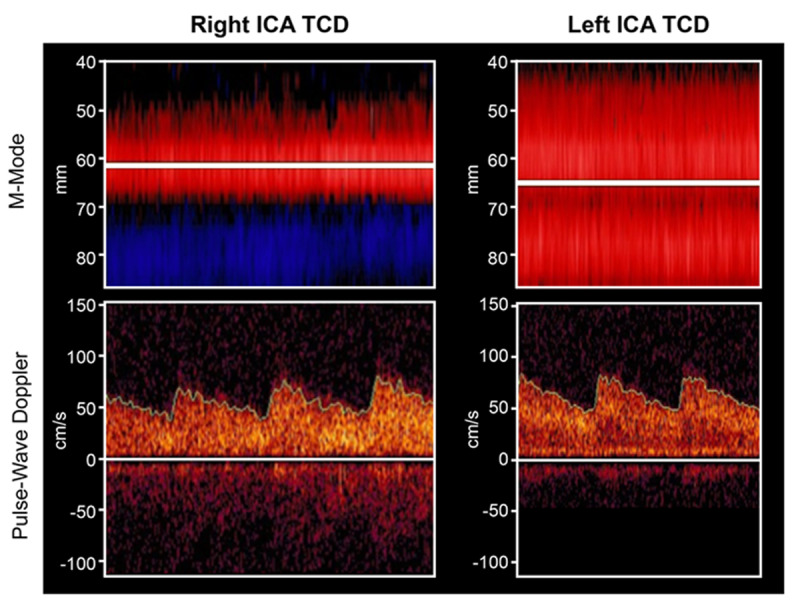
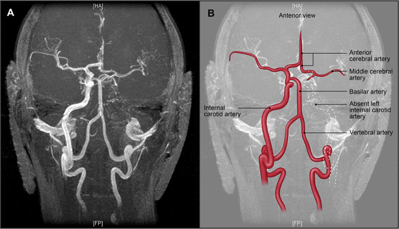
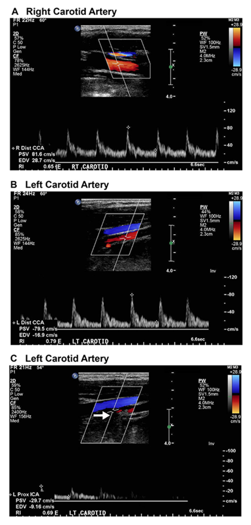
Agenesis or hypoplasia of the internal carotid artery (ICA) may easily be confused with dissection or occlusion. We report a case of a 24-year-old female with complaint of acute left-hand hypoesthesia and a history of occasional intermittent numbness of her right hand with myoclonic jerking. Because previous imaging studies over 2 years were interpreted as occlusion of the left ICA secondary to carotid dissection, the treating physician had prescribed anticoagulant therapy. During transcranial Doppler (TCD) examination, the spectral waveform was unexpectedly normal, prompting a repeat review of all imaging due to the TCD results. Magnetic resonance angiography (MRA) revealed the same "flame-like" appearance of the ICA origin. Late-phase digital subtraction angiography showed a small caliber cervical ICA (occluded at the skull base). Computed tomography demonstrated absence of the carotid canal, confirming an absent intracranial portion of the ICA and establishing a correct diagnosis of left internal carotid hypoplasia. Vascular ultrasound and TCD examinations are noninvasive and inexpensive tools that can improve the interpretation and understanding of the clinical significance of other "static" radiographic tests (MRA, digital subtraction angiography ). An accurate diagnosis is essential to avoid risky, aggressive treatment, such as anticoagulation for an "absent" dissection.


 分享
分享
 求助内容:
求助内容: 应助结果提醒方式:
 扫码关注我们
