A. Mariscal Martínez , E. Iglesias Bravo , H. Peris Alvà , P. Rodríguez Martínez , M. Luna Tomás , I. Pascual Miguel , P. Puyalto de Pablo
{"title":"Mamografía con contraste y marcaje con semilla magnética para la detección de enfermedad residual en el cáncer de mama tras tratamiento neoadyuvante","authors":"A. Mariscal Martínez , E. Iglesias Bravo , H. Peris Alvà , P. Rodríguez Martínez , M. Luna Tomás , I. Pascual Miguel , P. Puyalto de Pablo","doi":"10.1016/j.rx.2024.04.003","DOIUrl":null,"url":null,"abstract":"<div><h3>Purpose</h3><p>Assess whether contrast-enhanced mammography (CEM) enables an evaluation of the residual size of breast tumours following neoadjuvant systemic therapy (NAST) in patients initially marked with magnetic seed.</p></div><div><h3>Materials and methods</h3><p>This single-centre prospective study was performed between March 2022 and April 2023 with patients with invasive breast carcinoma and lesional marking with magnetic seed. CEM was performed before and after NAST. The lesion size in CEM after NAST was compared to the pathological examination after surgery. Differences between sizes were evaluated and we determined the diagnostic capability indices.</p></div><div><h3>Results</h3><p>The breast lesions marked with magnetic seed were successfully localised in the preoperative stage for the 42 patients included in the study and selective surgical excision was also achieved in all cases. Tumour diameter after NAST was determined by comparing enhancement on combined CEM images from before and after NAST. The mean diameter was 13.6 mm while post-surgical pathological examination determined the mean diameter to be 12.9 mm. There were therefore no statistically significant differences between the measurements.</p></div><div><h3>Conclusions</h3><p>There is a positive correlation and similarity between CEM and pathological examination with regards to the detection of residual disease after NAST, with high specificity and positive predictive value.</p></div>","PeriodicalId":31509,"journal":{"name":"RADIOLOGIA","volume":"66 5","pages":"Pages 419-430"},"PeriodicalIF":1.1000,"publicationDate":"2024-09-01","publicationTypes":"Journal Article","fieldsOfStudy":null,"isOpenAccess":false,"openAccessPdf":"","citationCount":"0","resultStr":null,"platform":"Semanticscholar","paperid":null,"PeriodicalName":"RADIOLOGIA","FirstCategoryId":"1085","ListUrlMain":"https://www.sciencedirect.com/science/article/pii/S0033833824000638","RegionNum":0,"RegionCategory":null,"ArticlePicture":[],"TitleCN":null,"AbstractTextCN":null,"PMCID":null,"EPubDate":"2024/5/30 0:00:00","PubModel":"Epub","JCR":"Q3","JCRName":"RADIOLOGY, NUCLEAR MEDICINE & MEDICAL IMAGING","Score":null,"Total":0}
引用次数: 0
Abstract
Purpose
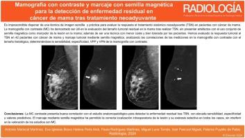
Assess whether contrast-enhanced mammography (CEM) enables an evaluation of the residual size of breast tumours following neoadjuvant systemic therapy (NAST) in patients initially marked with magnetic seed.
Materials and methods
This single-centre prospective study was performed between March 2022 and April 2023 with patients with invasive breast carcinoma and lesional marking with magnetic seed. CEM was performed before and after NAST. The lesion size in CEM after NAST was compared to the pathological examination after surgery. Differences between sizes were evaluated and we determined the diagnostic capability indices.
Results
The breast lesions marked with magnetic seed were successfully localised in the preoperative stage for the 42 patients included in the study and selective surgical excision was also achieved in all cases. Tumour diameter after NAST was determined by comparing enhancement on combined CEM images from before and after NAST. The mean diameter was 13.6 mm while post-surgical pathological examination determined the mean diameter to be 12.9 mm. There were therefore no statistically significant differences between the measurements.
Conclusions
There is a positive correlation and similarity between CEM and pathological examination with regards to the detection of residual disease after NAST, with high specificity and positive predictive value.
RADIOLOGIARADIOLOGY, NUCLEAR MEDICINE & MEDICAL IMAGING-
CiteScore
1.60
自引率
7.70%
发文量
105
审稿时长
52 days
期刊介绍:
La mejor revista para conocer de primera mano los originales más relevantes en la especialidad y las revisiones, casos y notas clínicas de mayor interés profesional. Además es la Publicación Oficial de la Sociedad Española de Radiología Médica.


 分享
分享
 求助内容:
求助内容: 应助结果提醒方式:
 扫码关注我们
