Dajeong Nam , Jaejung Park , Jaehong Lee , Juyoung Son , Ja-Eun Kim
{"title":"mTOR potentiates senescent phenotypes and primary cilia formation after cisplatin-induced G2 arrest in retinal pigment epithelial cells","authors":"Dajeong Nam , Jaejung Park , Jaehong Lee , Juyoung Son , Ja-Eun Kim","doi":"10.1016/j.cellsig.2024.111402","DOIUrl":null,"url":null,"abstract":"<div><p>Cisplatin, a platinum-based anticancer drug, is used to treat several types of cancer. Despite its effectiveness, cisplatin-induced side effects have often been reported. Although cisplatin-induced toxicities, such as apoptosis and/or necrosis, have been well studied, the fate of cells after exposure to sublethal doses of cisplatin needs further elucidation. Treatment with a sublethal dose of cisplatin induced cell cycle arrest at the G2 phase in retinal pigment epithelial cells. Following cisplatin withdrawal, the cells irreversibly exited the cell cycle and became senescent. Notably, the progression from the G2 to the G1 phase occurred without mitotic entry, a phenomenon referred to as mitotic bypass, resulting in the accumulation of cells containing 4N DNA content. Cisplatin-exposed cells exhibited morphological changes associated with senescence, including an enlarged size of cell and nucleus and increased granularity. In addition, the senescent cells possessed primary cilia and persistent DNA lesions. Senescence induced by transient exposure to cisplatin involves mTOR activation. Although transient co-exposure with an mTORC1 inhibitor rapamycin did not prevent mitotic bypass and entry into senescence, it delayed the progression of senescence and attenuated senescent phenotypes, resulting in shorter primary cilia formation. Conclusively, cisplatin induces senescence in retinal pigment epithelial cells by promoting mTOR activation.</p></div>","PeriodicalId":9902,"journal":{"name":"Cellular signalling","volume":"124 ","pages":"Article 111402"},"PeriodicalIF":3.7000,"publicationDate":"2024-09-07","publicationTypes":"Journal Article","fieldsOfStudy":null,"isOpenAccess":false,"openAccessPdf":"https://www.sciencedirect.com/science/article/pii/S089865682400370X/pdfft?md5=d45190ff7d660909042fd3d885037e81&pid=1-s2.0-S089865682400370X-main.pdf","citationCount":"0","resultStr":null,"platform":"Semanticscholar","paperid":null,"PeriodicalName":"Cellular signalling","FirstCategoryId":"99","ListUrlMain":"https://www.sciencedirect.com/science/article/pii/S089865682400370X","RegionNum":2,"RegionCategory":"生物学","ArticlePicture":[],"TitleCN":null,"AbstractTextCN":null,"PMCID":null,"EPubDate":"","PubModel":"","JCR":"Q2","JCRName":"CELL BIOLOGY","Score":null,"Total":0}
引用次数: 0
Abstract
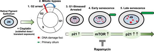
Cisplatin, a platinum-based anticancer drug, is used to treat several types of cancer. Despite its effectiveness, cisplatin-induced side effects have often been reported. Although cisplatin-induced toxicities, such as apoptosis and/or necrosis, have been well studied, the fate of cells after exposure to sublethal doses of cisplatin needs further elucidation. Treatment with a sublethal dose of cisplatin induced cell cycle arrest at the G2 phase in retinal pigment epithelial cells. Following cisplatin withdrawal, the cells irreversibly exited the cell cycle and became senescent. Notably, the progression from the G2 to the G1 phase occurred without mitotic entry, a phenomenon referred to as mitotic bypass, resulting in the accumulation of cells containing 4N DNA content. Cisplatin-exposed cells exhibited morphological changes associated with senescence, including an enlarged size of cell and nucleus and increased granularity. In addition, the senescent cells possessed primary cilia and persistent DNA lesions. Senescence induced by transient exposure to cisplatin involves mTOR activation. Although transient co-exposure with an mTORC1 inhibitor rapamycin did not prevent mitotic bypass and entry into senescence, it delayed the progression of senescence and attenuated senescent phenotypes, resulting in shorter primary cilia formation. Conclusively, cisplatin induces senescence in retinal pigment epithelial cells by promoting mTOR activation.
期刊介绍:
Cellular Signalling publishes original research describing fundamental and clinical findings on the mechanisms, actions and structural components of cellular signalling systems in vitro and in vivo.
Cellular Signalling aims at full length research papers defining signalling systems ranging from microorganisms to cells, tissues and higher organisms.


 分享
分享
 求助内容:
求助内容: 应助结果提醒方式:
 扫码关注我们
