Meagan Wu, Benjamin B. Massenburg, Dillan F. Villavisanis, Jinggang J. Ng, Dominic J. Romeo, Connor S. Wagner, Scott P. Bartlett, Jordan W. Swanson, Jesse A. Taylor
{"title":"Long-term photogrammetric outcomes of midface advancement in Apert syndrome: are we nearing normal?","authors":"Meagan Wu, Benjamin B. Massenburg, Dillan F. Villavisanis, Jinggang J. Ng, Dominic J. Romeo, Connor S. Wagner, Scott P. Bartlett, Jordan W. Swanson, Jesse A. Taylor","doi":"10.1007/s00381-024-06611-5","DOIUrl":null,"url":null,"abstract":"<h3 data-test=\"abstract-sub-heading\">Background</h3><p>The aesthetic goals of midface surgery in Apert syndrome are to correct the multi-planar midface deficiency and normalize facial ratios. This study characterizes the long-term photogrammetric outcomes of midface advancement in Apert syndrome.</p><h3 data-test=\"abstract-sub-heading\">Methods</h3><p>Patients with Apert syndrome who underwent midface distraction from 2000 to 2023 were retrospectively reviewed. Soft tissue measurements were applied to preoperative (T0), short-term postoperative (T1), and long-term postoperative (T2) photographs. Long-term changes were compared between subcranial and transcranial procedures, segmental and non-segmental osteotomies, and individual techniques. Frontal facial dimensions at time T2 were compared to age- and sex-matched normal controls from the NIMH-ChEFS face database.</p><h3 data-test=\"abstract-sub-heading\">Results</h3><p>Twenty patients had postoperative follow-up of T1 = 0.6 ± 0.4 and T2 = 4.7 ± 1.1 years and were compared to 40 normal controls. From time T0 to T2, middle facial third height increased (26.1 ± 3.0% to 27.8 ± 2.6%, <i>p</i> = 0.026), canthal tilt improved (− 7.6° ± 3.7° to − 3.9° ± 4.4°, <i>p</i> < 0.001), and facial convexity increased (182.9° ± 6.2° to 167.9° ± 8.6°, <i>p</i> < 0.001). From time T1 to T2, facial convexity flattened (159.4° ± 10.1° to 167.9° ± 8.6°, <i>p</i> < 0.001). Compared to controls, patients at time T2 had shorter middle facial third height (27.8 ± 2.6% vs. 32.0 ± 1.9%, <i>p</i> < 0.001) and reverse canthal tilt (− 3.9° ± 4.4° vs. 5.4° ± 2.6°, <i>p</i> < 0.001).</p><h3 data-test=\"abstract-sub-heading\">Conclusions</h3><p>A tailored treatment approach to the Apert midface yields varying degrees of resolution of central midfacial shortening, canthal tilt, and facial concavity at 5 years postoperatively. A slight reduction in facial convexity over time, likely reflecting pseudorelapse in the setting of sagittal overcorrection, should be anticipated. Greater utilization of segmental osteotomies may bring facial third ratios and canthal tilt closer to normal morphometric values.</p>","PeriodicalId":9970,"journal":{"name":"Child's Nervous System","volume":"27 1","pages":""},"PeriodicalIF":1.2000,"publicationDate":"2024-09-13","publicationTypes":"Journal Article","fieldsOfStudy":null,"isOpenAccess":false,"openAccessPdf":"","citationCount":"0","resultStr":null,"platform":"Semanticscholar","paperid":null,"PeriodicalName":"Child's Nervous System","FirstCategoryId":"3","ListUrlMain":"https://doi.org/10.1007/s00381-024-06611-5","RegionNum":4,"RegionCategory":"医学","ArticlePicture":[],"TitleCN":null,"AbstractTextCN":null,"PMCID":null,"EPubDate":"","PubModel":"","JCR":"Q4","JCRName":"CLINICAL NEUROLOGY","Score":null,"Total":0}
引用次数: 0
Abstract
Background
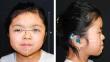
The aesthetic goals of midface surgery in Apert syndrome are to correct the multi-planar midface deficiency and normalize facial ratios. This study characterizes the long-term photogrammetric outcomes of midface advancement in Apert syndrome.
Methods
Patients with Apert syndrome who underwent midface distraction from 2000 to 2023 were retrospectively reviewed. Soft tissue measurements were applied to preoperative (T0), short-term postoperative (T1), and long-term postoperative (T2) photographs. Long-term changes were compared between subcranial and transcranial procedures, segmental and non-segmental osteotomies, and individual techniques. Frontal facial dimensions at time T2 were compared to age- and sex-matched normal controls from the NIMH-ChEFS face database.
Results
Twenty patients had postoperative follow-up of T1 = 0.6 ± 0.4 and T2 = 4.7 ± 1.1 years and were compared to 40 normal controls. From time T0 to T2, middle facial third height increased (26.1 ± 3.0% to 27.8 ± 2.6%, p = 0.026), canthal tilt improved (− 7.6° ± 3.7° to − 3.9° ± 4.4°, p < 0.001), and facial convexity increased (182.9° ± 6.2° to 167.9° ± 8.6°, p < 0.001). From time T1 to T2, facial convexity flattened (159.4° ± 10.1° to 167.9° ± 8.6°, p < 0.001). Compared to controls, patients at time T2 had shorter middle facial third height (27.8 ± 2.6% vs. 32.0 ± 1.9%, p < 0.001) and reverse canthal tilt (− 3.9° ± 4.4° vs. 5.4° ± 2.6°, p < 0.001).
Conclusions
A tailored treatment approach to the Apert midface yields varying degrees of resolution of central midfacial shortening, canthal tilt, and facial concavity at 5 years postoperatively. A slight reduction in facial convexity over time, likely reflecting pseudorelapse in the setting of sagittal overcorrection, should be anticipated. Greater utilization of segmental osteotomies may bring facial third ratios and canthal tilt closer to normal morphometric values.
期刊介绍:
The journal has been expanded to encompass all aspects of pediatric neurosciences concerning the developmental and acquired abnormalities of the nervous system and its coverings, functional disorders, epilepsy, spasticity, basic and clinical neuro-oncology, rehabilitation and trauma. Global pediatric neurosurgery is an additional field of interest that will be considered for publication in the journal.


 分享
分享
 求助内容:
求助内容: 应助结果提醒方式:
 扫码关注我们
