Yuan Ren, Jie Yang, Barbara Fujita, Yongli Zhang, Julien Berro
{"title":"Cross-regulations of two connected domains form a mechanical circuit for steady force transmission during clathrin-mediated endocytosis","authors":"Yuan Ren, Jie Yang, Barbara Fujita, Yongli Zhang, Julien Berro","doi":"10.1016/j.celrep.2024.114725","DOIUrl":null,"url":null,"abstract":"<p>Mechanical forces are transmitted from the actin cytoskeleton to the membrane during clathrin-mediated endocytosis (CME) in the fission yeast <em>Schizosaccharomyces pombe</em>. End4p directly transmits force in CME by binding to both the membrane (through the AP180 N-terminal homology [ANTH] domain) and F-actin (through the talin-HIP1/R/Sla2p actin-tethering C-terminal homology [THATCH] domain). We show that 7 pN force is required for stable binding between THATCH and F-actin. We also characterized a domain in End4p, Rend (rod domain in End4p), that resembles R12 of talin. Membrane localization of Rend primes the binding of THATCH to F-actin, and force-induced unfolding of Rend at 15 pN terminates the transmission of force. We show that the mechanical properties (mechanical stability, unfolding extension, hysteresis) of Rend and THATCH are tuned to form a circuit for the initiation, transmission, and termination of force between the actin cytoskeleton and membrane. The mechanical circuit by Rend and THATCH may be conserved and coopted evolutionarily in cell adhesion complexes.</p>","PeriodicalId":9798,"journal":{"name":"Cell reports","volume":"12 1 1","pages":""},"PeriodicalIF":6.9000,"publicationDate":"2024-09-13","publicationTypes":"Journal Article","fieldsOfStudy":null,"isOpenAccess":false,"openAccessPdf":"","citationCount":"0","resultStr":null,"platform":"Semanticscholar","paperid":null,"PeriodicalName":"Cell reports","FirstCategoryId":"99","ListUrlMain":"https://doi.org/10.1016/j.celrep.2024.114725","RegionNum":1,"RegionCategory":"生物学","ArticlePicture":[],"TitleCN":null,"AbstractTextCN":null,"PMCID":null,"EPubDate":"","PubModel":"","JCR":"Q1","JCRName":"CELL BIOLOGY","Score":null,"Total":0}
引用次数: 0
Abstract
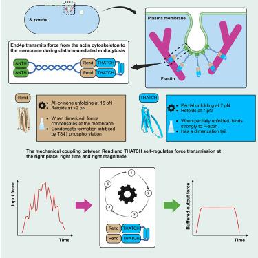
Mechanical forces are transmitted from the actin cytoskeleton to the membrane during clathrin-mediated endocytosis (CME) in the fission yeast Schizosaccharomyces pombe. End4p directly transmits force in CME by binding to both the membrane (through the AP180 N-terminal homology [ANTH] domain) and F-actin (through the talin-HIP1/R/Sla2p actin-tethering C-terminal homology [THATCH] domain). We show that 7 pN force is required for stable binding between THATCH and F-actin. We also characterized a domain in End4p, Rend (rod domain in End4p), that resembles R12 of talin. Membrane localization of Rend primes the binding of THATCH to F-actin, and force-induced unfolding of Rend at 15 pN terminates the transmission of force. We show that the mechanical properties (mechanical stability, unfolding extension, hysteresis) of Rend and THATCH are tuned to form a circuit for the initiation, transmission, and termination of force between the actin cytoskeleton and membrane. The mechanical circuit by Rend and THATCH may be conserved and coopted evolutionarily in cell adhesion complexes.
期刊介绍:
Cell Reports publishes high-quality research across the life sciences and focuses on new biological insight as its primary criterion for publication. The journal offers three primary article types: Reports, which are shorter single-point articles, research articles, which are longer and provide deeper mechanistic insights, and resources, which highlight significant technical advances or major informational datasets that contribute to biological advances. Reviews covering recent literature in emerging and active fields are also accepted.
The Cell Reports Portfolio includes gold open-access journals that cover life, medical, and physical sciences, and its mission is to make cutting-edge research and methodologies available to a wide readership.
The journal's professional in-house editors work closely with authors, reviewers, and the scientific advisory board, which consists of current and future leaders in their respective fields. The advisory board guides the scope, content, and quality of the journal, but editorial decisions are independently made by the in-house scientific editors of Cell Reports.


 分享
分享
 求助内容:
求助内容: 应助结果提醒方式:
 扫码关注我们
