Mena Shenouda, Eyjólfur Gudmundsson, Feng Li, Christopher M. Straus, Hedy L. Kindler, Arkadiusz Z. Dudek, Thomas Stinchcombe, Xiaofei Wang, Adam Starkey, Samuel G. Armato III
{"title":"Convolutional Neural Networks for Segmentation of Pleural Mesothelioma: Analysis of Probability Map Thresholds (CALGB 30901, Alliance)","authors":"Mena Shenouda, Eyjólfur Gudmundsson, Feng Li, Christopher M. Straus, Hedy L. Kindler, Arkadiusz Z. Dudek, Thomas Stinchcombe, Xiaofei Wang, Adam Starkey, Samuel G. Armato III","doi":"10.1007/s10278-024-01092-z","DOIUrl":null,"url":null,"abstract":"<p>The purpose of this study was to evaluate the impact of probability map threshold on pleural mesothelioma (PM) tumor delineations generated using a convolutional neural network (CNN). One hundred eighty-six CT scans from 48 PM patients were segmented by a VGG16/U-Net CNN. A radiologist modified the contours generated at a 0.5 probability threshold. Percent difference of tumor volume and overlap using the Dice Similarity Coefficient (DSC) were compared between the reference standard provided by the radiologist and CNN outputs for thresholds ranging from 0.001 to 0.9. CNN-derived contours consistently yielded smaller tumor volumes than radiologist contours. Reducing the probability threshold from 0.5 to 0.01 decreased the absolute percent volume difference, on average, from 42.93% to 26.60%. Median and mean DSC ranged from 0.57 to 0.59, with a peak at a threshold of 0.2; no distinct threshold was found for percent volume difference. The CNN exhibited deficiencies with specific disease presentations, such as severe pleural effusion or disease in the pleural fissure. No single output threshold in the CNN probability maps was optimal for both tumor volume and DSC. This study emphasized the importance of considering both figures of merit when evaluating deep learning-based tumor segmentations across probability thresholds. This work underscores the need to simultaneously assess tumor volume and spatial overlap when evaluating CNN performance. While automated segmentations may yield comparable tumor volumes to that of the reference standard, the spatial region delineated by the CNN at a specific threshold is equally important.</p>","PeriodicalId":50214,"journal":{"name":"Journal of Digital Imaging","volume":"3 1","pages":""},"PeriodicalIF":3.8000,"publicationDate":"2024-09-12","publicationTypes":"Journal Article","fieldsOfStudy":null,"isOpenAccess":false,"openAccessPdf":"","citationCount":"0","resultStr":null,"platform":"Semanticscholar","paperid":null,"PeriodicalName":"Journal of Digital Imaging","FirstCategoryId":"5","ListUrlMain":"https://doi.org/10.1007/s10278-024-01092-z","RegionNum":2,"RegionCategory":"工程技术","ArticlePicture":[],"TitleCN":null,"AbstractTextCN":null,"PMCID":null,"EPubDate":"","PubModel":"","JCR":"Q2","JCRName":"RADIOLOGY, NUCLEAR MEDICINE & MEDICAL IMAGING","Score":null,"Total":0}
引用次数: 0
Abstract
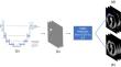
The purpose of this study was to evaluate the impact of probability map threshold on pleural mesothelioma (PM) tumor delineations generated using a convolutional neural network (CNN). One hundred eighty-six CT scans from 48 PM patients were segmented by a VGG16/U-Net CNN. A radiologist modified the contours generated at a 0.5 probability threshold. Percent difference of tumor volume and overlap using the Dice Similarity Coefficient (DSC) were compared between the reference standard provided by the radiologist and CNN outputs for thresholds ranging from 0.001 to 0.9. CNN-derived contours consistently yielded smaller tumor volumes than radiologist contours. Reducing the probability threshold from 0.5 to 0.01 decreased the absolute percent volume difference, on average, from 42.93% to 26.60%. Median and mean DSC ranged from 0.57 to 0.59, with a peak at a threshold of 0.2; no distinct threshold was found for percent volume difference. The CNN exhibited deficiencies with specific disease presentations, such as severe pleural effusion or disease in the pleural fissure. No single output threshold in the CNN probability maps was optimal for both tumor volume and DSC. This study emphasized the importance of considering both figures of merit when evaluating deep learning-based tumor segmentations across probability thresholds. This work underscores the need to simultaneously assess tumor volume and spatial overlap when evaluating CNN performance. While automated segmentations may yield comparable tumor volumes to that of the reference standard, the spatial region delineated by the CNN at a specific threshold is equally important.
期刊介绍:
The Journal of Digital Imaging (JDI) is the official peer-reviewed journal of the Society for Imaging Informatics in Medicine (SIIM). JDI’s goal is to enhance the exchange of knowledge encompassed by the general topic of Imaging Informatics in Medicine such as research and practice in clinical, engineering, and information technologies and techniques in all medical imaging environments. JDI topics are of interest to researchers, developers, educators, physicians, and imaging informatics professionals.
Suggested Topics
PACS and component systems; imaging informatics for the enterprise; image-enabled electronic medical records; RIS and HIS; digital image acquisition; image processing; image data compression; 3D, visualization, and multimedia; speech recognition; computer-aided diagnosis; facilities design; imaging vocabularies and ontologies; Transforming the Radiological Interpretation Process (TRIP™); DICOM and other standards; workflow and process modeling and simulation; quality assurance; archive integrity and security; teleradiology; digital mammography; and radiological informatics education.


 分享
分享
 求助内容:
求助内容: 应助结果提醒方式:
 扫码关注我们
