{"title":"Development of STING probes and visualization of STING in multiple tumor types","authors":"Huanhuan Liu, Jia Liu, Yingxi Chen, Hongzhang Yang, Jianyang Fang, Xinying Zeng, Jingru Zhang, Shilan Peng, Yuanyuan Liang, Rongqiang Zhuang, Gang Liu, Xianzhong Zhang, Zhide Guo","doi":"10.1007/s00259-024-06919-z","DOIUrl":null,"url":null,"abstract":"<h3 data-test=\"abstract-sub-heading\">Purpose</h3><p>The stimulator of interferon genes (STING) is a critical component of the innate immune system and plays a pivotal role in tumor immunotherapy. Developing non-invasive in vivo diagnostic methods for visualizing STING is highly valuable for STING-related immunotherapy. This work aimed to build a noninvasive imaging platform that can dynamically and quantitatively monitor tumor STING expression.</p><h3 data-test=\"abstract-sub-heading\">Methods</h3><p>We investigated the in vivo positron emission tomography (PET) imaging of STING-expressing tumors (B16F10, MC38, and Panc02) with STING-targeted radioprobe ([<sup>18</sup>F]F-CRI<sub>1</sub>). The expression of STING in tumors was quantified, and correlation analysis was performed between these results and the outcomes of PET imaging. Furthermore, we optimized the structure of [<sup>18</sup>F]F-CRI<sub>n</sub> with polyethylene glycol (PEG) to improve the pharmacokinetic characteristics in vivo. A comprehensive comparison of the imaging and biodistribution results obtained with the optimized probes was conducted in the B16F10 tumors.</p><h3 data-test=\"abstract-sub-heading\">Results</h3><p>The PET imaging results showed that the uptake of [<sup>18</sup>F]F-CRI<sub>1</sub> in tumors was positively correlated with the expression of STING in tumors (<i>r</i> = 0.9184, <i>P</i> < 0.001 at 0.5 h). The lipophilicity of the optimized probes was significantly reduced. As a result of employing optimized probes, B16F10 tumor-bearing mice exhibited significantly improved tumor visualization in PET imaging, along with a marked reduction in retention within non-target areas such as the gallbladder and intestines. Biodistribution experiments further validated the efficacy of probe optimization in reducing uptake in non-target areas.</p><h3 data-test=\"abstract-sub-heading\">Conclusion</h3><p>In summary, this work demonstrated a promising pathway for the development of STING-targeted radioprobes, advancing in vivo PET imaging capabilities.</p><h3 data-test=\"abstract-sub-heading\">Graphical Abstract</h3>","PeriodicalId":11909,"journal":{"name":"European Journal of Nuclear Medicine and Molecular Imaging","volume":"78 1","pages":""},"PeriodicalIF":7.6000,"publicationDate":"2024-09-18","publicationTypes":"Journal Article","fieldsOfStudy":null,"isOpenAccess":false,"openAccessPdf":"","citationCount":"0","resultStr":null,"platform":"Semanticscholar","paperid":null,"PeriodicalName":"European Journal of Nuclear Medicine and Molecular Imaging","FirstCategoryId":"3","ListUrlMain":"https://doi.org/10.1007/s00259-024-06919-z","RegionNum":1,"RegionCategory":"医学","ArticlePicture":[],"TitleCN":null,"AbstractTextCN":null,"PMCID":null,"EPubDate":"","PubModel":"","JCR":"Q1","JCRName":"RADIOLOGY, NUCLEAR MEDICINE & MEDICAL IMAGING","Score":null,"Total":0}
引用次数: 0
Abstract
Purpose
The stimulator of interferon genes (STING) is a critical component of the innate immune system and plays a pivotal role in tumor immunotherapy. Developing non-invasive in vivo diagnostic methods for visualizing STING is highly valuable for STING-related immunotherapy. This work aimed to build a noninvasive imaging platform that can dynamically and quantitatively monitor tumor STING expression.
Methods
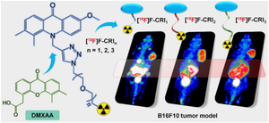
We investigated the in vivo positron emission tomography (PET) imaging of STING-expressing tumors (B16F10, MC38, and Panc02) with STING-targeted radioprobe ([18F]F-CRI1). The expression of STING in tumors was quantified, and correlation analysis was performed between these results and the outcomes of PET imaging. Furthermore, we optimized the structure of [18F]F-CRIn with polyethylene glycol (PEG) to improve the pharmacokinetic characteristics in vivo. A comprehensive comparison of the imaging and biodistribution results obtained with the optimized probes was conducted in the B16F10 tumors.
Results
The PET imaging results showed that the uptake of [18F]F-CRI1 in tumors was positively correlated with the expression of STING in tumors (r = 0.9184, P < 0.001 at 0.5 h). The lipophilicity of the optimized probes was significantly reduced. As a result of employing optimized probes, B16F10 tumor-bearing mice exhibited significantly improved tumor visualization in PET imaging, along with a marked reduction in retention within non-target areas such as the gallbladder and intestines. Biodistribution experiments further validated the efficacy of probe optimization in reducing uptake in non-target areas.
Conclusion
In summary, this work demonstrated a promising pathway for the development of STING-targeted radioprobes, advancing in vivo PET imaging capabilities.
期刊介绍:
The European Journal of Nuclear Medicine and Molecular Imaging serves as a platform for the exchange of clinical and scientific information within nuclear medicine and related professions. It welcomes international submissions from professionals involved in the functional, metabolic, and molecular investigation of diseases. The journal's coverage spans physics, dosimetry, radiation biology, radiochemistry, and pharmacy, providing high-quality peer review by experts in the field. Known for highly cited and downloaded articles, it ensures global visibility for research work and is part of the EJNMMI journal family.


 分享
分享

 求助内容:
求助内容: 应助结果提醒方式:
 扫码关注我们
