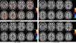
{"title":"Disrupted brain functional asymmetry at rest in patients with major depressive disorder associated with sleep disturbances","authors":"Dan Lv, Yangpan Ou, Huabing Li, Feng Liu, Ping Li, Dongsheng Lv, Jingping Zhao, Wenbin Guo","doi":"10.1007/s11682-024-00924-4","DOIUrl":null,"url":null,"abstract":"<p>Sleep disturbances (SD) are common in major depressive disorder (MDD) patients. Brain functional asymmetry is crucial for understanding MDD pathophysiology. Previous studies using the parameter of asymmetry (PAS) approach have found brain functional asymmetry disruption in MDD. However, this has not been explored in MDD patients with SD. This study examined 26 MDD patients with SD, 34 MDD patients without SD, and 34 healthy controls using resting-state functional magnetic resonance imaging scans. SD symptoms were quantified using the 17-item Hamilton Rating Scale for Depression. PAS approach was used to evaluate functional asymmetry. MDD patients with SD displayed increased PAS in the left middle frontal gyrus (MFG)/inferior frontal gyrus (IFG) and decreased PAS in the left parahippocampal gyrus (PHG) compared to MDD patients without SD. Increased PAS in the left MFG/IFG was positively correlated with SD severity, and a negative correlation was found between decreased PAS in the left PHG and SD scores in all MDD patients. Receiver operating characteristic analysis indicated that increased PAS in the left MFG/IFG and decreased PAS in the left PHG may serve as potential neuroimaging markers to differentiate MDD patients with SD from those without SD with Area Under Curve values of 0.8157 and 0.8068, respectively. These results highlighted that increased PAS in the left MFG/IFG and decreased PAS in the left PHG may be considered a prominent feature associated with SD symptoms of MDD patients, potentially serving as imaging markers to discriminate between MDD patients with and without SD.</p>","PeriodicalId":9192,"journal":{"name":"Brain Imaging and Behavior","volume":"40 1","pages":""},"PeriodicalIF":2.4000,"publicationDate":"2024-09-14","publicationTypes":"Journal Article","fieldsOfStudy":null,"isOpenAccess":false,"openAccessPdf":"","citationCount":"0","resultStr":null,"platform":"Semanticscholar","paperid":null,"PeriodicalName":"Brain Imaging and Behavior","FirstCategoryId":"3","ListUrlMain":"https://doi.org/10.1007/s11682-024-00924-4","RegionNum":3,"RegionCategory":"医学","ArticlePicture":[],"TitleCN":null,"AbstractTextCN":null,"PMCID":null,"EPubDate":"","PubModel":"","JCR":"Q2","JCRName":"NEUROIMAGING","Score":null,"Total":0}
引用次数: 0
Abstract
Sleep disturbances (SD) are common in major depressive disorder (MDD) patients. Brain functional asymmetry is crucial for understanding MDD pathophysiology. Previous studies using the parameter of asymmetry (PAS) approach have found brain functional asymmetry disruption in MDD. However, this has not been explored in MDD patients with SD. This study examined 26 MDD patients with SD, 34 MDD patients without SD, and 34 healthy controls using resting-state functional magnetic resonance imaging scans. SD symptoms were quantified using the 17-item Hamilton Rating Scale for Depression. PAS approach was used to evaluate functional asymmetry. MDD patients with SD displayed increased PAS in the left middle frontal gyrus (MFG)/inferior frontal gyrus (IFG) and decreased PAS in the left parahippocampal gyrus (PHG) compared to MDD patients without SD. Increased PAS in the left MFG/IFG was positively correlated with SD severity, and a negative correlation was found between decreased PAS in the left PHG and SD scores in all MDD patients. Receiver operating characteristic analysis indicated that increased PAS in the left MFG/IFG and decreased PAS in the left PHG may serve as potential neuroimaging markers to differentiate MDD patients with SD from those without SD with Area Under Curve values of 0.8157 and 0.8068, respectively. These results highlighted that increased PAS in the left MFG/IFG and decreased PAS in the left PHG may be considered a prominent feature associated with SD symptoms of MDD patients, potentially serving as imaging markers to discriminate between MDD patients with and without SD.
期刊介绍:
Brain Imaging and Behavior is a bi-monthly, peer-reviewed journal, that publishes clinically relevant research using neuroimaging approaches to enhance our understanding of disorders of higher brain function. The journal is targeted at clinicians and researchers in fields concerned with human brain-behavior relationships, such as neuropsychology, psychiatry, neurology, neurosurgery, rehabilitation, and cognitive neuroscience.


 分享
分享
 求助内容:
求助内容: 应助结果提醒方式:
 扫码关注我们
