Simultaneous labial and lingual augmented corticotomy assisted presurgical orthodontics in class III patients: the morphological aspects of the mandibular anterior ridge with cone-beam computed tomography
Jian Liu, Yi Li, Yu Fu, Xiaotong Li, Xiao Xu, Li Xu, Jianxia Hou
{"title":"Simultaneous labial and lingual augmented corticotomy assisted presurgical orthodontics in class III patients: the morphological aspects of the mandibular anterior ridge with cone-beam computed tomography","authors":"Jian Liu, Yi Li, Yu Fu, Xiaotong Li, Xiao Xu, Li Xu, Jianxia Hou","doi":"10.1007/s00784-024-05805-0","DOIUrl":null,"url":null,"abstract":"<h3 data-test=\"abstract-sub-heading\">Objectives</h3><p>This study aims to investigate the changes in alveolar bone following the simultaneous performance of labial and lingual augmented corticotomy (LLAC) in patients with insufficient alveolar bone thickness on both the labial and lingual sides of the mandibular anterior teeth during presurgical orthodontic treatment.</p><h3 data-test=\"abstract-sub-heading\">Materials and methods</h3><p>Thirth-five surgical patients with skeletal Class III malocclusion were included: 19 (LLAC group) accepted LLAC surgery during presurgical orthodontic treatment, and 16 (non-surgery group, NS) accepted traditional presurgical orthodontic treatment. Cone-beam computed tomography (CBCT) scans were obtained before treatment (T0) and at the completion of presurgical orthodontic treatment (T1). The amount of vertical alveolar bone and contour area of the alveolar bone in the labial and lingual sides of mandibular incisors were measured.</p><h3 data-test=\"abstract-sub-heading\">Results</h3><p>After presurgical orthodontic treatment, the contour area of the alveolar bone at each level on the lingual side and alveolar bone level on both sides decreased significantly in the NS group (<i>P</i> < 0.001). However, the labial and lingual bone contour area at each level and bone level increased significantly in the LLAC group (<i>P</i> < 0.001). The bone formation rate in the lingual apical region was the highest, significantly different from other sites (<i>P</i> < 0.001).</p><h3 data-test=\"abstract-sub-heading\">Conclusions</h3><p>During presurgical orthodontic treatment, LLAC can significantly increase the contour area of the labio-lingual alveolar bone in the mandibular anterior teeth to facilitate safe and effective orthodontic decompensation in skeletal Class III patients.</p><h3 data-test=\"abstract-sub-heading\">Clinical relevance</h3><p>: This surgery has positive clinical significance in patients lacking bone thickness (< 0.5 mm) in the labial and lingual sides of the lower incisors.</p>","PeriodicalId":10461,"journal":{"name":"Clinical Oral Investigations","volume":"18 1","pages":""},"PeriodicalIF":3.1000,"publicationDate":"2024-09-17","publicationTypes":"Journal Article","fieldsOfStudy":null,"isOpenAccess":false,"openAccessPdf":"","citationCount":"0","resultStr":null,"platform":"Semanticscholar","paperid":null,"PeriodicalName":"Clinical Oral Investigations","FirstCategoryId":"3","ListUrlMain":"https://doi.org/10.1007/s00784-024-05805-0","RegionNum":2,"RegionCategory":"医学","ArticlePicture":[],"TitleCN":null,"AbstractTextCN":null,"PMCID":null,"EPubDate":"","PubModel":"","JCR":"Q1","JCRName":"DENTISTRY, ORAL SURGERY & MEDICINE","Score":null,"Total":0}
引用次数: 0
Abstract
Objectives
This study aims to investigate the changes in alveolar bone following the simultaneous performance of labial and lingual augmented corticotomy (LLAC) in patients with insufficient alveolar bone thickness on both the labial and lingual sides of the mandibular anterior teeth during presurgical orthodontic treatment.
Materials and methods
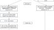
Thirth-five surgical patients with skeletal Class III malocclusion were included: 19 (LLAC group) accepted LLAC surgery during presurgical orthodontic treatment, and 16 (non-surgery group, NS) accepted traditional presurgical orthodontic treatment. Cone-beam computed tomography (CBCT) scans were obtained before treatment (T0) and at the completion of presurgical orthodontic treatment (T1). The amount of vertical alveolar bone and contour area of the alveolar bone in the labial and lingual sides of mandibular incisors were measured.
Results
After presurgical orthodontic treatment, the contour area of the alveolar bone at each level on the lingual side and alveolar bone level on both sides decreased significantly in the NS group (P < 0.001). However, the labial and lingual bone contour area at each level and bone level increased significantly in the LLAC group (P < 0.001). The bone formation rate in the lingual apical region was the highest, significantly different from other sites (P < 0.001).
Conclusions
During presurgical orthodontic treatment, LLAC can significantly increase the contour area of the labio-lingual alveolar bone in the mandibular anterior teeth to facilitate safe and effective orthodontic decompensation in skeletal Class III patients.
Clinical relevance
: This surgery has positive clinical significance in patients lacking bone thickness (< 0.5 mm) in the labial and lingual sides of the lower incisors.
期刊介绍:
The journal Clinical Oral Investigations is a multidisciplinary, international forum for publication of research from all fields of oral medicine. The journal publishes original scientific articles and invited reviews which provide up-to-date results of basic and clinical studies in oral and maxillofacial science and medicine. The aim is to clarify the relevance of new results to modern practice, for an international readership. Coverage includes maxillofacial and oral surgery, prosthetics and restorative dentistry, operative dentistry, endodontics, periodontology, orthodontics, dental materials science, clinical trials, epidemiology, pedodontics, oral implant, preventive dentistiry, oral pathology, oral basic sciences and more.


 分享
分享
 求助内容:
求助内容: 应助结果提醒方式:
 扫码关注我们
