{"title":"Correlation Between Swallowing and Cognitive Brain Networks in Older Adults via Functional MRI.","authors":"Jie Wang, Mengqing Zhang, Hongmei Wen, Cheng Yang, Xiaomei Wei, Yonghui Wang, Zulin Dou","doi":"10.1111/joor.13855","DOIUrl":null,"url":null,"abstract":"<p><strong>Background: </strong>Clinical evidence supports that swallowing function is correlated with cognition, but the neurobiological mechanism associated with cognitive impairment and dysphagia remains unclear.</p><p><strong>Objectives: </strong>To compare the brain activation patterns of the swallowing and the cognitive tasks and explore neural associations between swallowing and cognitive function via task-related functional magnetic resonance imaging (fMRI).</p><p><strong>Methods: </strong>A total of 13 healthy older adults (aged > 60 years) were recruited. Participants underwent the clinical dementia rating (CDR) test and the Montreal Cognitive Assessment (MoCA). A block-designed task-related fMRI study was conducted where each participant completed both swallowing and cognitive tasks within a single session. During the swallowing task, participants swallowed 2 mL of thickened water, while the Stroop Colour Word Test (SCWT) served as the cognitive task. First-level analysis of swallowing time-series images utilised the general linear model (GLM) with Statistical Parametric Mapping (SPM), applying a voxel threshold of p < 0.001 for significance. Common activations in brain regions during swallowing and cognitive tasks were extracted at the group level, with significance set at p < 0.05, corrected for multiple comparisons using the false discovery rate (FDR), with a minimum cluster size of 20 voxels. Correlation analysis between behavioural measurements and imaging signals was also conducted.</p><p><strong>Results: </strong>Some regions were commonly activated in both task networks; these regions were the bilateral occipital lobe, cerebellum, lingual gyrus, fusiform, middle frontal gyrus, precentral and postcentral gyrus, right supramarginal and inferior parietal lobe. Most importantly, the average beta value of cognitive and swallowing tasks in these areas are both significantly negative related to the MoCA score. Furthermore, opposite signal changes were seen in the bilateral prefrontal lobes during the swallowing task, while positive activation in the bilateral prefrontal lobes was observed during the SCWT. Postcentral gyrus activation was more extensive than precentral gyrus activation in the swallowing task.</p><p><strong>Conclusion: </strong>The common activation of swallowing and cognitive tasks had multiple foci. The activity of cognitive and swallowing task in these areas is significantly negative correlated with the MoCA score. These findings may help to illustrate the association between dysphagia and cognitive impairment due to the common brain regions involved in cognition and swallowing and may provide a reference for further rehabilitation of dysphagia.</p><p><strong>Trial registration: </strong>Clinical Trial: (Chinese Clinical Trial Registry): ChiCTR1900021795.</p>","PeriodicalId":16605,"journal":{"name":"Journal of oral rehabilitation","volume":null,"pages":null},"PeriodicalIF":3.1000,"publicationDate":"2024-12-01","publicationTypes":"Journal Article","fieldsOfStudy":null,"isOpenAccess":false,"openAccessPdf":"","citationCount":"0","resultStr":null,"platform":"Semanticscholar","paperid":null,"PeriodicalName":"Journal of oral rehabilitation","FirstCategoryId":"3","ListUrlMain":"https://doi.org/10.1111/joor.13855","RegionNum":3,"RegionCategory":"医学","ArticlePicture":[],"TitleCN":null,"AbstractTextCN":null,"PMCID":null,"EPubDate":"2024/9/20 0:00:00","PubModel":"Epub","JCR":"Q1","JCRName":"DENTISTRY, ORAL SURGERY & MEDICINE","Score":null,"Total":0}
引用次数: 0
Abstract
Background: Clinical evidence supports that swallowing function is correlated with cognition, but the neurobiological mechanism associated with cognitive impairment and dysphagia remains unclear.
Objectives: To compare the brain activation patterns of the swallowing and the cognitive tasks and explore neural associations between swallowing and cognitive function via task-related functional magnetic resonance imaging (fMRI).
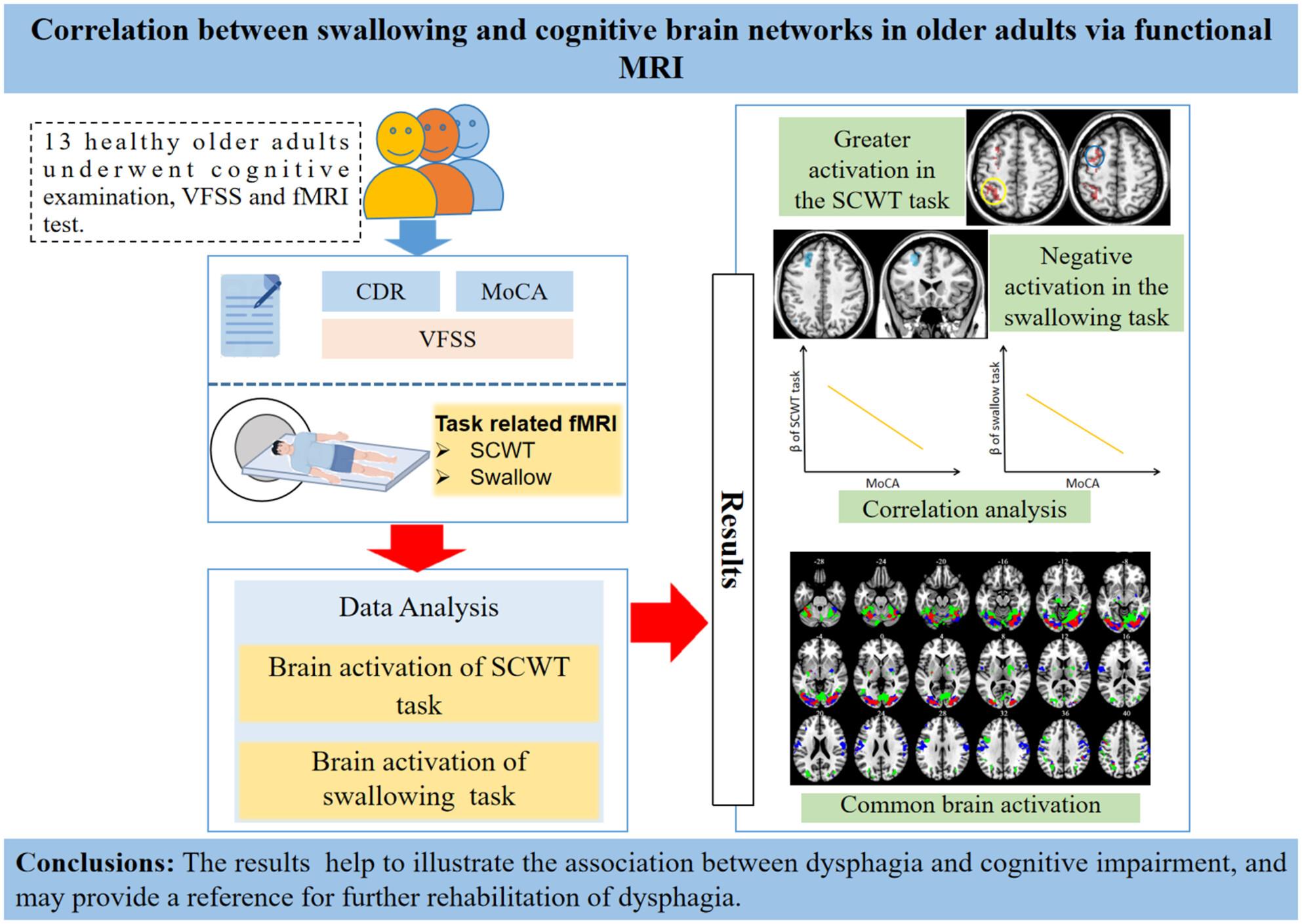
Methods: A total of 13 healthy older adults (aged > 60 years) were recruited. Participants underwent the clinical dementia rating (CDR) test and the Montreal Cognitive Assessment (MoCA). A block-designed task-related fMRI study was conducted where each participant completed both swallowing and cognitive tasks within a single session. During the swallowing task, participants swallowed 2 mL of thickened water, while the Stroop Colour Word Test (SCWT) served as the cognitive task. First-level analysis of swallowing time-series images utilised the general linear model (GLM) with Statistical Parametric Mapping (SPM), applying a voxel threshold of p < 0.001 for significance. Common activations in brain regions during swallowing and cognitive tasks were extracted at the group level, with significance set at p < 0.05, corrected for multiple comparisons using the false discovery rate (FDR), with a minimum cluster size of 20 voxels. Correlation analysis between behavioural measurements and imaging signals was also conducted.
Results: Some regions were commonly activated in both task networks; these regions were the bilateral occipital lobe, cerebellum, lingual gyrus, fusiform, middle frontal gyrus, precentral and postcentral gyrus, right supramarginal and inferior parietal lobe. Most importantly, the average beta value of cognitive and swallowing tasks in these areas are both significantly negative related to the MoCA score. Furthermore, opposite signal changes were seen in the bilateral prefrontal lobes during the swallowing task, while positive activation in the bilateral prefrontal lobes was observed during the SCWT. Postcentral gyrus activation was more extensive than precentral gyrus activation in the swallowing task.
Conclusion: The common activation of swallowing and cognitive tasks had multiple foci. The activity of cognitive and swallowing task in these areas is significantly negative correlated with the MoCA score. These findings may help to illustrate the association between dysphagia and cognitive impairment due to the common brain regions involved in cognition and swallowing and may provide a reference for further rehabilitation of dysphagia.
期刊介绍:
Journal of Oral Rehabilitation aims to be the most prestigious journal of dental research within all aspects of oral rehabilitation and applied oral physiology. It covers all diagnostic and clinical management aspects necessary to re-establish a subjective and objective harmonious oral function.
Oral rehabilitation may become necessary as a result of developmental or acquired disturbances in the orofacial region, orofacial traumas, or a variety of dental and oral diseases (primarily dental caries and periodontal diseases) and orofacial pain conditions. As such, oral rehabilitation in the twenty-first century is a matter of skilful diagnosis and minimal, appropriate intervention, the nature of which is intimately linked to a profound knowledge of oral physiology, oral biology, and dental and oral pathology.
The scientific content of the journal therefore strives to reflect the best of evidence-based clinical dentistry. Modern clinical management should be based on solid scientific evidence gathered about diagnostic procedures and the properties and efficacy of the chosen intervention (e.g. material science, biological, toxicological, pharmacological or psychological aspects). The content of the journal also reflects documentation of the possible side-effects of rehabilitation, and includes prognostic perspectives of the treatment modalities chosen.