Amedeo Piazza, Sergio Corvino, Daniel Ballesteros, Alice Campeggi, Edoardo Agosti, Simona Serioli, Francesco Corrivetti, Carlo Bortolotti, Matteo De Notaris
{"title":"Neuroanatomical photogrammetric models using smartphones: a comparison of apps","authors":"Amedeo Piazza, Sergio Corvino, Daniel Ballesteros, Alice Campeggi, Edoardo Agosti, Simona Serioli, Francesco Corrivetti, Carlo Bortolotti, Matteo De Notaris","doi":"10.1007/s00701-024-06264-y","DOIUrl":null,"url":null,"abstract":"<p>A deep knowledge of the surgical anatomy of the target area is mandatory for a successful operative procedure. For this purpose, over the years, many teaching and learning methods have been described, from the most ancient cadaveric dissection to the most recent virtual reality, each with their respective pros and cons. Photogrammetry, an emergent technique, allows for the creation of three-dimensional (3D) models and reconstructions. Thanks to the spreading of photogrammetry nowadays it is possible to generate these models using professional software or even smartphone apps. This study aims to compare the neuroanatomical photogrammetric models generated by the two most utilized smartphone applications in this domain, Metascan and 3D-Scanner, through quantitative analysis.</p><p>Two human head specimens (four sides) were examined. Anatomical dissection was segmented into five stages to systematically expose well-defined structures. After each stage, a photogrammetric model was generated using two prominent smartphone applications. These models were then subjected to both quantitative and qualitative analysis, with a specific focus on comparing the mesh density as a measure of model resolution and accuracy. Appropriate consent was obtained for the publication of the cadaver's image.</p><p>The quantitative analysis revealed that the models generated by Metascan app consistently demonstrated superior mesh density compared to those from 3D-Scanner, indicating a higher level of detail and potential for precise anatomical representation.</p><p>Enabling depth perception, capturing high-quality images, offering flexibility in viewpoints: photogrammetry provides researchers with unprecedented opportunities to explore and understand the intricate and magnificent structure of the brain. However, it is of paramount importance to develop and apply rigorous quality control systems to ensure data integrity and reliability of findings in neurological research. This study has demonstrated the superiority of Metascan in processing photogrammetric models for neuroanatomical studies.</p>","PeriodicalId":7370,"journal":{"name":"Acta Neurochirurgica","volume":"166 1","pages":""},"PeriodicalIF":1.9000,"publicationDate":"2024-09-24","publicationTypes":"Journal Article","fieldsOfStudy":null,"isOpenAccess":false,"openAccessPdf":"https://www.ncbi.nlm.nih.gov/pmc/articles/PMC11422470/pdf/","citationCount":"0","resultStr":null,"platform":"Semanticscholar","paperid":null,"PeriodicalName":"Acta Neurochirurgica","FirstCategoryId":"3","ListUrlMain":"https://link.springer.com/article/10.1007/s00701-024-06264-y","RegionNum":3,"RegionCategory":"医学","ArticlePicture":[],"TitleCN":null,"AbstractTextCN":null,"PMCID":null,"EPubDate":"","PubModel":"","JCR":"Q3","JCRName":"CLINICAL NEUROLOGY","Score":null,"Total":0}
引用次数: 0
Abstract
A deep knowledge of the surgical anatomy of the target area is mandatory for a successful operative procedure. For this purpose, over the years, many teaching and learning methods have been described, from the most ancient cadaveric dissection to the most recent virtual reality, each with their respective pros and cons. Photogrammetry, an emergent technique, allows for the creation of three-dimensional (3D) models and reconstructions. Thanks to the spreading of photogrammetry nowadays it is possible to generate these models using professional software or even smartphone apps. This study aims to compare the neuroanatomical photogrammetric models generated by the two most utilized smartphone applications in this domain, Metascan and 3D-Scanner, through quantitative analysis.
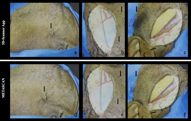
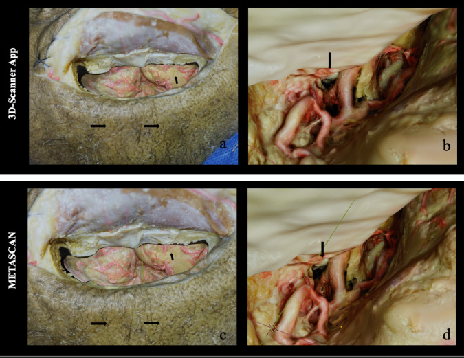
Two human head specimens (four sides) were examined. Anatomical dissection was segmented into five stages to systematically expose well-defined structures. After each stage, a photogrammetric model was generated using two prominent smartphone applications. These models were then subjected to both quantitative and qualitative analysis, with a specific focus on comparing the mesh density as a measure of model resolution and accuracy. Appropriate consent was obtained for the publication of the cadaver's image.
The quantitative analysis revealed that the models generated by Metascan app consistently demonstrated superior mesh density compared to those from 3D-Scanner, indicating a higher level of detail and potential for precise anatomical representation.
Enabling depth perception, capturing high-quality images, offering flexibility in viewpoints: photogrammetry provides researchers with unprecedented opportunities to explore and understand the intricate and magnificent structure of the brain. However, it is of paramount importance to develop and apply rigorous quality control systems to ensure data integrity and reliability of findings in neurological research. This study has demonstrated the superiority of Metascan in processing photogrammetric models for neuroanatomical studies.
期刊介绍:
The journal "Acta Neurochirurgica" publishes only original papers useful both to research and clinical work. Papers should deal with clinical neurosurgery - diagnosis and diagnostic techniques, operative surgery and results, postoperative treatment - or with research work in neuroscience if the underlying questions or the results are of neurosurgical interest. Reports on congresses are given in brief accounts. As official organ of the European Association of Neurosurgical Societies the journal publishes all announcements of the E.A.N.S. and reports on the activities of its member societies. Only contributions written in English will be accepted.


 分享
分享
 求助内容:
求助内容: 应助结果提醒方式:
 扫码关注我们
