{"title":"A longitudinal MRI analysis reveals altered brain connectivity and microstructural changes in a transgenic mouse model of Alzheimer's disease","authors":"Ricardo Magalhães , Fernanda Marques , Erwan Selingue , Fawzi Boumezbeur , Sébastien Mériaux , Nuno Sousa","doi":"10.1016/j.nbd.2024.106679","DOIUrl":null,"url":null,"abstract":"<div><div>Alzheimer's disease (AD) is characterized by progressive cognitive decline and neuropathological changes, yet the underlying neurobiological mechanisms remain elusive. Here, we employed a multimodal longitudinal neuroimaging approach, using anatomical and functional sequences on a high field magnetic resonance imaging (MRI) preclinical scanner, to investigate alterations in brain connectivity and white matter microstructure in a transgenic mouse model of AD (J20) when compared to wild-type (WT) littermates. Functional connectivity analysis revealed distinct network disruptions in J20 mice, primarily involving connections between posterior and anterior brain regions; importantly, a significant interaction between group and age highlighted an exacerbation of these connectivity changes with advancing age in J20 mice. In addition, significant reductions in fractional anisotropy (FA) were observed in the corpus callosum of J20 mice compared to WT, indicative of microstructural alterations consistent with white matter pathology. The observed alterations in brain connectivity and microstructure provide valuable insights into the spatiotemporal processes underlying AD-related decline and underscore the utility of multimodal neuroimaging in elucidating the neurobiological substrates of AD pathology in animal models.</div></div>","PeriodicalId":19097,"journal":{"name":"Neurobiology of Disease","volume":"201 ","pages":"Article 106679"},"PeriodicalIF":5.6000,"publicationDate":"2024-10-15","publicationTypes":"Journal Article","fieldsOfStudy":null,"isOpenAccess":false,"openAccessPdf":"https://www.sciencedirect.com/science/article/pii/S0969996124002791/pdfft?md5=43450098de98d9cbb9aa5700ef7ec892&pid=1-s2.0-S0969996124002791-main.pdf","citationCount":"0","resultStr":null,"platform":"Semanticscholar","paperid":null,"PeriodicalName":"Neurobiology of Disease","FirstCategoryId":"3","ListUrlMain":"https://www.sciencedirect.com/science/article/pii/S0969996124002791","RegionNum":2,"RegionCategory":"医学","ArticlePicture":[],"TitleCN":null,"AbstractTextCN":null,"PMCID":null,"EPubDate":"2024/9/23 0:00:00","PubModel":"Epub","JCR":"Q1","JCRName":"NEUROSCIENCES","Score":null,"Total":0}
引用次数: 0
Abstract
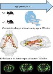
Alzheimer's disease (AD) is characterized by progressive cognitive decline and neuropathological changes, yet the underlying neurobiological mechanisms remain elusive. Here, we employed a multimodal longitudinal neuroimaging approach, using anatomical and functional sequences on a high field magnetic resonance imaging (MRI) preclinical scanner, to investigate alterations in brain connectivity and white matter microstructure in a transgenic mouse model of AD (J20) when compared to wild-type (WT) littermates. Functional connectivity analysis revealed distinct network disruptions in J20 mice, primarily involving connections between posterior and anterior brain regions; importantly, a significant interaction between group and age highlighted an exacerbation of these connectivity changes with advancing age in J20 mice. In addition, significant reductions in fractional anisotropy (FA) were observed in the corpus callosum of J20 mice compared to WT, indicative of microstructural alterations consistent with white matter pathology. The observed alterations in brain connectivity and microstructure provide valuable insights into the spatiotemporal processes underlying AD-related decline and underscore the utility of multimodal neuroimaging in elucidating the neurobiological substrates of AD pathology in animal models.
期刊介绍:
Neurobiology of Disease is a major international journal at the interface between basic and clinical neuroscience. The journal provides a forum for the publication of top quality research papers on: molecular and cellular definitions of disease mechanisms, the neural systems and underpinning behavioral disorders, the genetics of inherited neurological and psychiatric diseases, nervous system aging, and findings relevant to the development of new therapies.


 分享
分享
 求助内容:
求助内容: 应助结果提醒方式:
 扫码关注我们
