Zachary P. Christensen, Edward G. Freedman, John J. Foxe
{"title":"Autism is associated with in vivo changes in gray matter neurite architecture","authors":"Zachary P. Christensen, Edward G. Freedman, John J. Foxe","doi":"10.1002/aur.3239","DOIUrl":null,"url":null,"abstract":"<p>Postmortem investigations in autism have identified anomalies in neural cytoarchitecture across limbic, cerebellar, and neocortical networks. These anomalies include narrow cell mini-columns and variable neuron density. However, difficulty obtaining sufficient post-mortem samples has often prevented investigations from converging on reproducible measures. Recent advances in processing magnetic resonance diffusion weighted images (DWI) make in vivo characterization of neuronal cytoarchitecture a potential alternative to post-mortem studies. Using extensive DWI data from the Adolescent Brain Cognitive Development<sup>sm</sup> (ABCD®) study 142 individuals with an autism diagnosis were compared with 8971 controls using a restriction spectrum imaging (RSI) framework that characterized total neurite density (TND), its component restricted normalized directional diffusion (RND), and restricted normalized isotropic diffusion (RNI). A significant decrease in TND was observed in autism in the right cerebellar cortex (<i>β</i> = −0.005, SE =0.0015, <i>p</i> = 0.0267), with significant decreases in RNI and significant increases in RND found diffusely throughout posterior and anterior aspects of the brain, respectively. Furthermore, these regions remained significant in <i>post-hoc</i> analysis when the autism sample was compared against a subset of 1404 individuals with other psychiatric conditions (pulled from the original 8971). These findings highlight the importance of characterizing neuron cytoarchitecture in autism and the significance of their incorporation as physiological covariates in future studies.</p>","PeriodicalId":131,"journal":{"name":"Autism Research","volume":"17 11","pages":"2261-2277"},"PeriodicalIF":5.6000,"publicationDate":"2024-09-26","publicationTypes":"Journal Article","fieldsOfStudy":null,"isOpenAccess":false,"openAccessPdf":"https://onlinelibrary.wiley.com/doi/epdf/10.1002/aur.3239","citationCount":"0","resultStr":null,"platform":"Semanticscholar","paperid":null,"PeriodicalName":"Autism Research","FirstCategoryId":"3","ListUrlMain":"https://onlinelibrary.wiley.com/doi/10.1002/aur.3239","RegionNum":2,"RegionCategory":"医学","ArticlePicture":[],"TitleCN":null,"AbstractTextCN":null,"PMCID":null,"EPubDate":"","PubModel":"","JCR":"Q1","JCRName":"BEHAVIORAL SCIENCES","Score":null,"Total":0}
引用次数: 0
Abstract
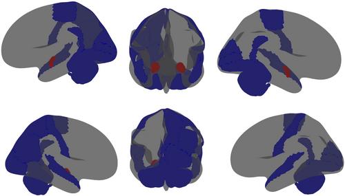
Postmortem investigations in autism have identified anomalies in neural cytoarchitecture across limbic, cerebellar, and neocortical networks. These anomalies include narrow cell mini-columns and variable neuron density. However, difficulty obtaining sufficient post-mortem samples has often prevented investigations from converging on reproducible measures. Recent advances in processing magnetic resonance diffusion weighted images (DWI) make in vivo characterization of neuronal cytoarchitecture a potential alternative to post-mortem studies. Using extensive DWI data from the Adolescent Brain Cognitive Developmentsm (ABCD®) study 142 individuals with an autism diagnosis were compared with 8971 controls using a restriction spectrum imaging (RSI) framework that characterized total neurite density (TND), its component restricted normalized directional diffusion (RND), and restricted normalized isotropic diffusion (RNI). A significant decrease in TND was observed in autism in the right cerebellar cortex (β = −0.005, SE =0.0015, p = 0.0267), with significant decreases in RNI and significant increases in RND found diffusely throughout posterior and anterior aspects of the brain, respectively. Furthermore, these regions remained significant in post-hoc analysis when the autism sample was compared against a subset of 1404 individuals with other psychiatric conditions (pulled from the original 8971). These findings highlight the importance of characterizing neuron cytoarchitecture in autism and the significance of their incorporation as physiological covariates in future studies.
期刊介绍:
AUTISM RESEARCH will cover the developmental disorders known as Pervasive Developmental Disorders (or autism spectrum disorders – ASDs). The Journal focuses on basic genetic, neurobiological and psychological mechanisms and how these influence developmental processes in ASDs.


 分享
分享
 求助内容:
求助内容: 应助结果提醒方式:
 扫码关注我们
