{"title":"Alterations in anterior lens capsule structure and LTBP-2 expression in primary angle-closure glaucoma.","authors":"Xiaofeng Tian, Liyun Yuan, Liangpin Li, Xiaoyong Yuan","doi":"10.1136/bmjophth-2023-001535","DOIUrl":null,"url":null,"abstract":"<p><strong>Objective: </strong>This study investigated the role of latent-transforming growth factor β-binding protein 2 (LTBP-2) in primary angle-closure glaucoma (PACG) by analysing its expression and the ultrastructure of the anterior lens capsule in PACG patients with age-related cataract (ARC).</p><p><strong>Methods: </strong>Tissue samples of the anterior lens capsule were collected from patients undergoing cataract phacoemulsification surgery. Patients in the experimental group were diagnosed with primary angle-closure (PAC) combined with ARC (PAC+ARC) and PACG combined with ARC (PACG+ARC). The control group consisted of patients with only ARC. The techniques used included scanning electron microscopy, real-time fluorescence quantitative polymerase chain reaction (RT-qPCR), western blotting and immunofluorescence.</p><p><strong>Results: </strong>Ultrastructural analysis revealed disordered connections in PAC+ARC, loose connections in PACG+ARC and well-ordered connections in ARC. RT-qPCR and western blotting showed significantly lower LTBP-2 mRNA and protein expression in PAC+ARC and PACG+ARC than in ARC, with PAC+ARC having the lowest levels. Immunofluorescence confirmed these findings, showing varying LTBP-2 fluorescence intensities across groups.</p><p><strong>Conclusion: </strong>The study identified ultrastructural changes in the anterior lens capsules in PACG accompanied by reduced LTBP-2 expression, especially in PAC+ARC patients. This suggests a potential role for LTBP-2 in PACG development, warranting further investigation.</p>","PeriodicalId":9286,"journal":{"name":"BMJ Open Ophthalmology","volume":"9 1","pages":""},"PeriodicalIF":2.2000,"publicationDate":"2024-09-24","publicationTypes":"Journal Article","fieldsOfStudy":null,"isOpenAccess":false,"openAccessPdf":"https://www.ncbi.nlm.nih.gov/pmc/articles/PMC11423727/pdf/","citationCount":"0","resultStr":null,"platform":"Semanticscholar","paperid":null,"PeriodicalName":"BMJ Open Ophthalmology","FirstCategoryId":"1085","ListUrlMain":"https://doi.org/10.1136/bmjophth-2023-001535","RegionNum":0,"RegionCategory":null,"ArticlePicture":[],"TitleCN":null,"AbstractTextCN":null,"PMCID":null,"EPubDate":"","PubModel":"","JCR":"Q2","JCRName":"OPHTHALMOLOGY","Score":null,"Total":0}
引用次数: 0
Abstract
Objective: This study investigated the role of latent-transforming growth factor β-binding protein 2 (LTBP-2) in primary angle-closure glaucoma (PACG) by analysing its expression and the ultrastructure of the anterior lens capsule in PACG patients with age-related cataract (ARC).
Methods: Tissue samples of the anterior lens capsule were collected from patients undergoing cataract phacoemulsification surgery. Patients in the experimental group were diagnosed with primary angle-closure (PAC) combined with ARC (PAC+ARC) and PACG combined with ARC (PACG+ARC). The control group consisted of patients with only ARC. The techniques used included scanning electron microscopy, real-time fluorescence quantitative polymerase chain reaction (RT-qPCR), western blotting and immunofluorescence.
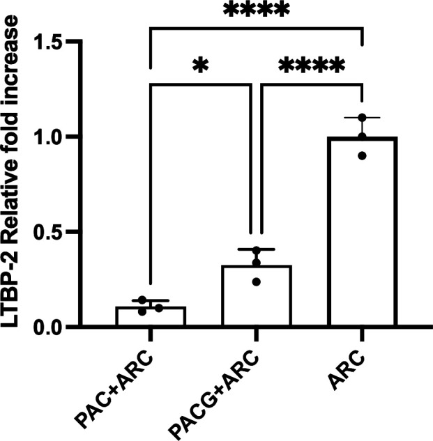
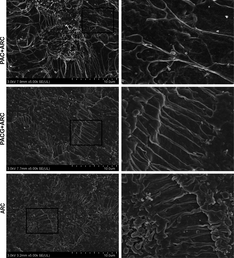
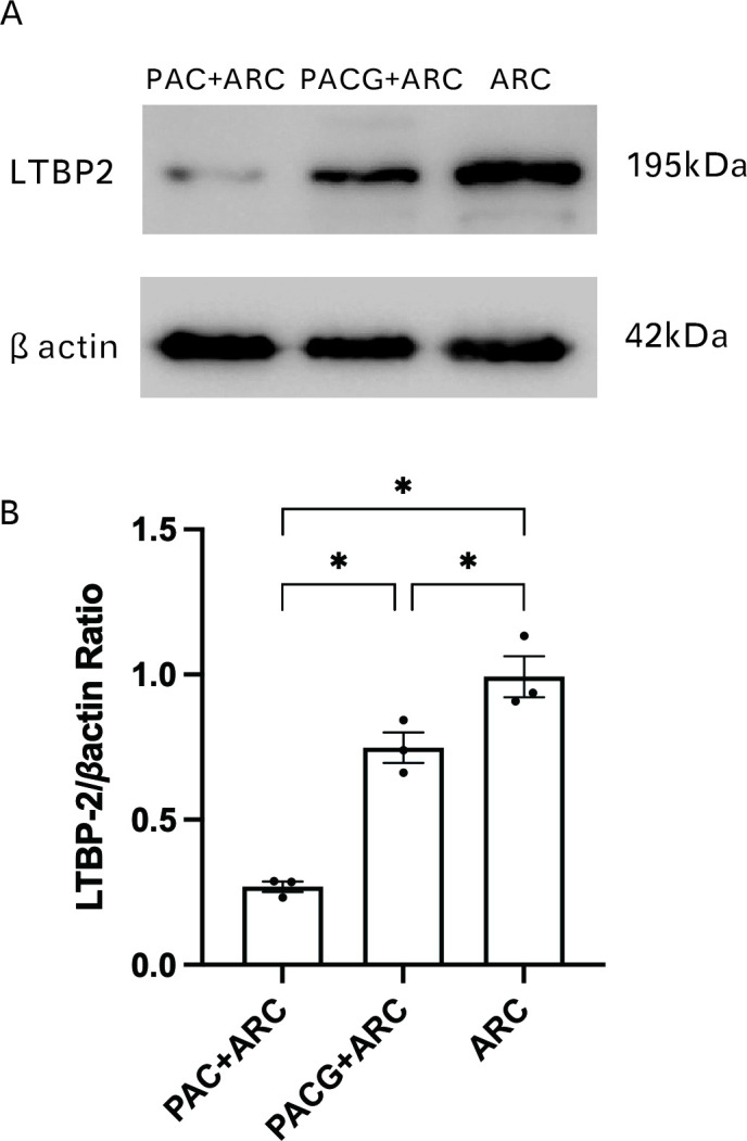
Results: Ultrastructural analysis revealed disordered connections in PAC+ARC, loose connections in PACG+ARC and well-ordered connections in ARC. RT-qPCR and western blotting showed significantly lower LTBP-2 mRNA and protein expression in PAC+ARC and PACG+ARC than in ARC, with PAC+ARC having the lowest levels. Immunofluorescence confirmed these findings, showing varying LTBP-2 fluorescence intensities across groups.
Conclusion: The study identified ultrastructural changes in the anterior lens capsules in PACG accompanied by reduced LTBP-2 expression, especially in PAC+ARC patients. This suggests a potential role for LTBP-2 in PACG development, warranting further investigation.


 分享
分享
 求助内容:
求助内容: 应助结果提醒方式:
 扫码关注我们
