{"title":"A case report of critical aortic stenosis diagnosed utilizing non-imaging continuous wave Doppler probe.","authors":"Edward D Shin, Eugene Fan","doi":"10.1093/ehjcr/ytae501","DOIUrl":null,"url":null,"abstract":"<p><strong>Background: </strong>Aortic stenosis (AS) is the most commonly acquired valvular pathology in the western world. Aortic stenosis severity is typically evaluated with Doppler echocardiography. Evaluation of aortic gradients using standard Doppler echocardiography from the apical window may underestimate the true gradient due to misalignment of blood flow to the ultrasound beam and is often better evaluated from other imaging windows using a non-imaging continuous wave Doppler (NI-CWD) probe. Herein, we describe a unique case of AS being underestimated by dynamic acoustic shadowing from the apical window rather than beam misalignment.</p><p><strong>Case summary: </strong>The patient is a Hispanic 31-year-old male with a congenital bicuspid aortic valve who underwent a balloon aortic valvuloplasty at age 13. At age 31, the patient underwent a repeat transthoracic echocardiogram (TTE) that was largely unchanged from his prior TTE from 15 years prior. Notably on this TTE, there was acoustic shadowing of colour Doppler in the distal left ventricular outflow tract and aortic valve during systole. While gradients only suggested moderate AS, the degree of left ventricular hypertrophy was suspicious for more severe AS. Only by using the NI-CWD probe at the right sternal border were we able to identify very severe AS with a peak velocity of 6.5 m/s and a mean gradient of 100 mmHg.</p><p><strong>Discussion: </strong>In our specific case, dynamic acoustic shadowing of the aortic valve from the apical window obscured both imaging and Doppler signals. This acoustic shadowing was not present from the right sternal border with the NI-CWD probe, leading to an over 100% increase in aortic valve peak velocity and proper correction of AS severity. This allowed for expedited care and underscores the importance of such techniques.</p>","PeriodicalId":11910,"journal":{"name":"European Heart Journal: Case Reports","volume":"8 10","pages":"ytae501"},"PeriodicalIF":0.8000,"publicationDate":"2024-09-14","publicationTypes":"Journal Article","fieldsOfStudy":null,"isOpenAccess":false,"openAccessPdf":"https://www.ncbi.nlm.nih.gov/pmc/articles/PMC11443958/pdf/","citationCount":"0","resultStr":null,"platform":"Semanticscholar","paperid":null,"PeriodicalName":"European Heart Journal: Case Reports","FirstCategoryId":"1085","ListUrlMain":"https://doi.org/10.1093/ehjcr/ytae501","RegionNum":0,"RegionCategory":null,"ArticlePicture":[],"TitleCN":null,"AbstractTextCN":null,"PMCID":null,"EPubDate":"2024/10/1 0:00:00","PubModel":"eCollection","JCR":"Q4","JCRName":"CARDIAC & CARDIOVASCULAR SYSTEMS","Score":null,"Total":0}
引用次数: 0
Abstract
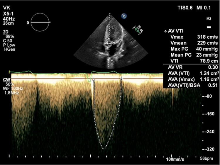
Background: Aortic stenosis (AS) is the most commonly acquired valvular pathology in the western world. Aortic stenosis severity is typically evaluated with Doppler echocardiography. Evaluation of aortic gradients using standard Doppler echocardiography from the apical window may underestimate the true gradient due to misalignment of blood flow to the ultrasound beam and is often better evaluated from other imaging windows using a non-imaging continuous wave Doppler (NI-CWD) probe. Herein, we describe a unique case of AS being underestimated by dynamic acoustic shadowing from the apical window rather than beam misalignment.
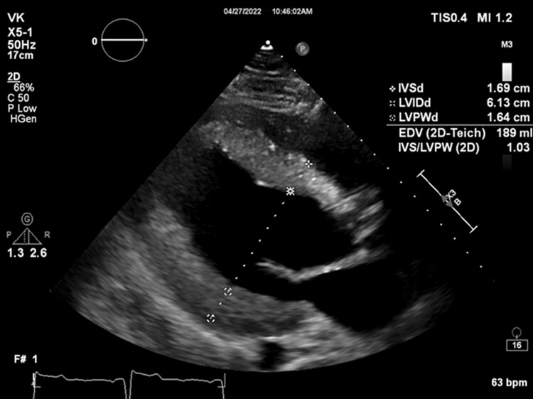
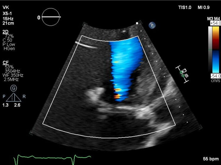
Case summary: The patient is a Hispanic 31-year-old male with a congenital bicuspid aortic valve who underwent a balloon aortic valvuloplasty at age 13. At age 31, the patient underwent a repeat transthoracic echocardiogram (TTE) that was largely unchanged from his prior TTE from 15 years prior. Notably on this TTE, there was acoustic shadowing of colour Doppler in the distal left ventricular outflow tract and aortic valve during systole. While gradients only suggested moderate AS, the degree of left ventricular hypertrophy was suspicious for more severe AS. Only by using the NI-CWD probe at the right sternal border were we able to identify very severe AS with a peak velocity of 6.5 m/s and a mean gradient of 100 mmHg.
Discussion: In our specific case, dynamic acoustic shadowing of the aortic valve from the apical window obscured both imaging and Doppler signals. This acoustic shadowing was not present from the right sternal border with the NI-CWD probe, leading to an over 100% increase in aortic valve peak velocity and proper correction of AS severity. This allowed for expedited care and underscores the importance of such techniques.


 分享
分享
 求助内容:
求助内容: 应助结果提醒方式:
 扫码关注我们
