Rahul Sreedasyam , Bryce G. Wilson , Patricia R. Ferrandez , Elliot L. Botvinick , Vasan Venugopalan
{"title":"An optical system for cellular mechanostimulation in 3D hydrogels","authors":"Rahul Sreedasyam , Bryce G. Wilson , Patricia R. Ferrandez , Elliot L. Botvinick , Vasan Venugopalan","doi":"10.1016/j.actbio.2024.09.050","DOIUrl":null,"url":null,"abstract":"<div><div>We introduce a method utilizing single laser-generated cavitation bubbles to stimulate cellular mechanotransduction in dermal fibroblasts embedded within 3D hydrogels. We demonstrate that fibroblasts embedded in either amorphous or fibrillar hydrogels engage in Ca<sup>2+</sup> signaling following exposure to an impulsive mechanical stimulus provided by a single 250 µm diameter laser-generated cavitation bubble. We find that the spatial extent of the cellular signaling is larger for cells embedded within a fibrous collagen hydrogel as compared to those embedded within an amorphous polyvinyl alcohol polymer (SLO-PVA) hydrogel. Additionally, for fibroblasts embedded in collagen, we find an increased range of cellular mechanosensitivity for cells that are polarized relative to the radial axis as compared to the circumferential axis. By contrast, fibroblasts embedded within SLO-PVA did not display orientation-dependent mechanosensitivity. Fibroblasts embedded in hydrogels and cultured in calcium-free media did not show cavitation-induced mechanotransduction; implicating calcium signaling based on transmembrane Ca<sup>2+</sup> transport. This study demonstrates the utility of single laser-generated cavitation bubbles to provide local non-invasive impulsive mechanical stimuli within 3D hydrogel tissue models with concurrent imaging using optical microscopy.</div></div><div><h3>Statement of significance</h3><div>Currently, there are limited methods for the non-invasive real-time assessment of cellular sensitivity to mechanical stimuli within 3D tissue scaffolds. We describe an original approach that utilizes a pulsed laser microbeam within a standard laser scanning microscope system to generate single cavitation bubbles to provide impulsive mechanostimulation to cells within 3D fibrillar and amorphous hydrogels. Using this technique, we measure the cellular mechanosensitivity of primary human dermal fibroblasts embedded in amorphous and fibrillar hydrogels, thereby providing a useful method to examine cellular mechanotransduction in 3D biomaterials. Moreover, the implementation of our method within a standard optical microscope makes it suitable for broad adoption by cellular mechanotransduction researchers and opens the possibility of high-throughput evaluation of biomaterials with respect to cellular mechanosignaling.</div></div>","PeriodicalId":237,"journal":{"name":"Acta Biomaterialia","volume":"189 ","pages":"Pages 439-448"},"PeriodicalIF":9.6000,"publicationDate":"2024-11-01","publicationTypes":"Journal Article","fieldsOfStudy":null,"isOpenAccess":false,"openAccessPdf":"","citationCount":"0","resultStr":null,"platform":"Semanticscholar","paperid":null,"PeriodicalName":"Acta Biomaterialia","FirstCategoryId":"5","ListUrlMain":"https://www.sciencedirect.com/science/article/pii/S1742706124005786","RegionNum":1,"RegionCategory":"医学","ArticlePicture":[],"TitleCN":null,"AbstractTextCN":null,"PMCID":null,"EPubDate":"2024/10/3 0:00:00","PubModel":"Epub","JCR":"Q1","JCRName":"ENGINEERING, BIOMEDICAL","Score":null,"Total":0}
引用次数: 0
Abstract
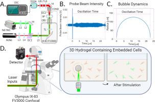
We introduce a method utilizing single laser-generated cavitation bubbles to stimulate cellular mechanotransduction in dermal fibroblasts embedded within 3D hydrogels. We demonstrate that fibroblasts embedded in either amorphous or fibrillar hydrogels engage in Ca2+ signaling following exposure to an impulsive mechanical stimulus provided by a single 250 µm diameter laser-generated cavitation bubble. We find that the spatial extent of the cellular signaling is larger for cells embedded within a fibrous collagen hydrogel as compared to those embedded within an amorphous polyvinyl alcohol polymer (SLO-PVA) hydrogel. Additionally, for fibroblasts embedded in collagen, we find an increased range of cellular mechanosensitivity for cells that are polarized relative to the radial axis as compared to the circumferential axis. By contrast, fibroblasts embedded within SLO-PVA did not display orientation-dependent mechanosensitivity. Fibroblasts embedded in hydrogels and cultured in calcium-free media did not show cavitation-induced mechanotransduction; implicating calcium signaling based on transmembrane Ca2+ transport. This study demonstrates the utility of single laser-generated cavitation bubbles to provide local non-invasive impulsive mechanical stimuli within 3D hydrogel tissue models with concurrent imaging using optical microscopy.
Statement of significance
Currently, there are limited methods for the non-invasive real-time assessment of cellular sensitivity to mechanical stimuli within 3D tissue scaffolds. We describe an original approach that utilizes a pulsed laser microbeam within a standard laser scanning microscope system to generate single cavitation bubbles to provide impulsive mechanostimulation to cells within 3D fibrillar and amorphous hydrogels. Using this technique, we measure the cellular mechanosensitivity of primary human dermal fibroblasts embedded in amorphous and fibrillar hydrogels, thereby providing a useful method to examine cellular mechanotransduction in 3D biomaterials. Moreover, the implementation of our method within a standard optical microscope makes it suitable for broad adoption by cellular mechanotransduction researchers and opens the possibility of high-throughput evaluation of biomaterials with respect to cellular mechanosignaling.
期刊介绍:
Acta Biomaterialia is a monthly peer-reviewed scientific journal published by Elsevier. The journal was established in January 2005. The editor-in-chief is W.R. Wagner (University of Pittsburgh). The journal covers research in biomaterials science, including the interrelationship of biomaterial structure and function from macroscale to nanoscale. Topical coverage includes biomedical and biocompatible materials.


 分享
分享
 求助内容:
求助内容: 应助结果提醒方式:
 扫码关注我们
