Comparative analysis of PDL1 and cluster of differentiation 68 marker expression in oral squamous cell carcinoma patients: Correlation with depth of invasion and immunofluorescence through immunohistochemistry.
Mehdi Shahabinejad, Siavash Bagheri Shirvan, Golnaz Fatemi, Farnaz Mohajer Tehran
{"title":"Comparative analysis of PDL1 and cluster of differentiation 68 marker expression in oral squamous cell carcinoma patients: Correlation with depth of invasion and immunofluorescence through immunohistochemistry.","authors":"Mehdi Shahabinejad, Siavash Bagheri Shirvan, Golnaz Fatemi, Farnaz Mohajer Tehran","doi":"","DOIUrl":null,"url":null,"abstract":"<p><strong>Background: </strong>Over the past 5 years, the use of immune checkpoint inhibitors in the treatment of head-and-neck squamous cell carcinoma (HNSCC) has increased. Both programmed death-ligand 1 (PD-L1) and cluster of differentiation 68 (CD68) are overexpressed in various carcinomas. Consequently, evaluating the expression of CD68 and PD-L1 in HNSCC lesions may lead to detecting a possible marker for HNSCC. This study aimed to evaluate the expression of PDL1 and CD68 markers in a patient with oral squamous cell carcinoma (OSCC) and examine its relationship with depth of invasion (DOI) and immunofluorescence (IF) through immunohistochemistry.</p><p><strong>Materials and methods: </strong>This cross-sectional study was conducted in the School of Dentistry, Mashhad University of Medical Sciences, Mashhad, Iran, Department of Oral and Maxillofacial Pathology. Thirty-four paraffin blocks and demographic information of 15 female and 19 male OSCC patients were collected. Following sample preparations, immunohistochemical staining was performed. Subsequently, each tissue section was analyzed for tumor-infiltrating lymphocytes by CD68 marker and PD-L1 expression. Data analysis was conducted using SPSS software (version 25). Chi-square, Shapiro-Wilk, and independent <i>t</i>-analytical tests were employed for statistical assessments. <i>P</i> < 0.05 was remarked as statistically significant.</p><p><strong>Results: </strong>CD68 and PDL1 expression in the squamous cell carcinoma (SCC) group was higher than the control group (<i>P</i> < 0.001). There was an increasing expression of PDL1 and CD68 as the grade of the disease progressed (<i>P</i> < 0.001 for each), as well as an increasing expression of IF and DOI.</p><p><strong>Conclusion: </strong>The expression levels of CD68 and PDL1 were elevated in SCC tissues in comparison to the unaffected, healthy parts of the tissue section.</p>","PeriodicalId":11016,"journal":{"name":"Dental Research Journal","volume":"21 ","pages":"48"},"PeriodicalIF":0.0000,"publicationDate":"2024-08-21","publicationTypes":"Journal Article","fieldsOfStudy":null,"isOpenAccess":false,"openAccessPdf":"https://www.ncbi.nlm.nih.gov/pmc/articles/PMC11457987/pdf/","citationCount":"0","resultStr":null,"platform":"Semanticscholar","paperid":null,"PeriodicalName":"Dental Research Journal","FirstCategoryId":"1085","ListUrlMain":"","RegionNum":0,"RegionCategory":null,"ArticlePicture":[],"TitleCN":null,"AbstractTextCN":null,"PMCID":null,"EPubDate":"2024/1/1 0:00:00","PubModel":"eCollection","JCR":"Q2","JCRName":"Dentistry","Score":null,"Total":0}
引用次数: 0
Abstract
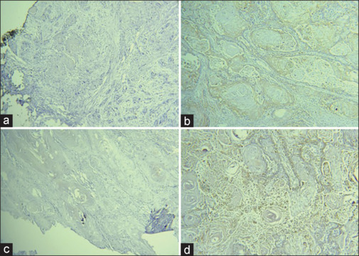
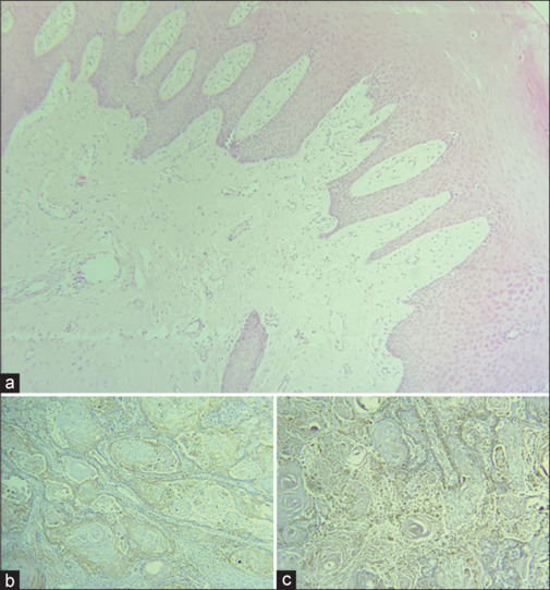
Background: Over the past 5 years, the use of immune checkpoint inhibitors in the treatment of head-and-neck squamous cell carcinoma (HNSCC) has increased. Both programmed death-ligand 1 (PD-L1) and cluster of differentiation 68 (CD68) are overexpressed in various carcinomas. Consequently, evaluating the expression of CD68 and PD-L1 in HNSCC lesions may lead to detecting a possible marker for HNSCC. This study aimed to evaluate the expression of PDL1 and CD68 markers in a patient with oral squamous cell carcinoma (OSCC) and examine its relationship with depth of invasion (DOI) and immunofluorescence (IF) through immunohistochemistry.
Materials and methods: This cross-sectional study was conducted in the School of Dentistry, Mashhad University of Medical Sciences, Mashhad, Iran, Department of Oral and Maxillofacial Pathology. Thirty-four paraffin blocks and demographic information of 15 female and 19 male OSCC patients were collected. Following sample preparations, immunohistochemical staining was performed. Subsequently, each tissue section was analyzed for tumor-infiltrating lymphocytes by CD68 marker and PD-L1 expression. Data analysis was conducted using SPSS software (version 25). Chi-square, Shapiro-Wilk, and independent t-analytical tests were employed for statistical assessments. P < 0.05 was remarked as statistically significant.
Results: CD68 and PDL1 expression in the squamous cell carcinoma (SCC) group was higher than the control group (P < 0.001). There was an increasing expression of PDL1 and CD68 as the grade of the disease progressed (P < 0.001 for each), as well as an increasing expression of IF and DOI.
Conclusion: The expression levels of CD68 and PDL1 were elevated in SCC tissues in comparison to the unaffected, healthy parts of the tissue section.
期刊介绍:
Dental Research Journal, a publication of Isfahan University of Medical Sciences, is a peer-reviewed online journal with Bimonthly print on demand compilation of issues published. The journal’s full text is available online at http://www.drjjournal.net. The journal allows free access (Open Access) to its contents and permits authors to self-archive final accepted version of the articles on any OAI-compliant institutional / subject-based repository. The journal will cover technical and clinical studies related to health, ethical and social issues in field of Dentistry. Articles with clinical interest and implications will be given preference.
 分享
分享


 求助内容:
求助内容: 应助结果提醒方式:
 扫码关注我们
