Kayla Le, Victoria Riggs, Seng Wai Yap, Martina Ernestova, Kimberley Sebastian
{"title":"Computed tomographic features of severe horn infection in a male Scimitar-horned oryx (Oryx dammah).","authors":"Kayla Le, Victoria Riggs, Seng Wai Yap, Martina Ernestova, Kimberley Sebastian","doi":"10.1111/vru.13440","DOIUrl":null,"url":null,"abstract":"<p><p>A 3-year-old male Scimitar-horned oryx presented for evaluation of an acutely deformed right horn with right head tilt and right facial pain. Computed tomographic evaluation revealed an increased volume of central fluid/soft tissue attenuation with gas-attenuating foci within the right horn. The right horn was amputated at the right horn base. Imaging and histopathologic features were consistent with emphysematous osteomyelitis. Following treatment, the patient returned to normal behavior. This is the first veterinary report describing the computed tomographic features of a horn infection in a Scimitar-horned oryx.</p>","PeriodicalId":23581,"journal":{"name":"Veterinary Radiology & Ultrasound","volume":" ","pages":"e13440"},"PeriodicalIF":1.5000,"publicationDate":"2025-01-01","publicationTypes":"Journal Article","fieldsOfStudy":null,"isOpenAccess":false,"openAccessPdf":"https://www.ncbi.nlm.nih.gov/pmc/articles/PMC11617606/pdf/","citationCount":"0","resultStr":null,"platform":"Semanticscholar","paperid":null,"PeriodicalName":"Veterinary Radiology & Ultrasound","FirstCategoryId":"97","ListUrlMain":"https://doi.org/10.1111/vru.13440","RegionNum":2,"RegionCategory":"农林科学","ArticlePicture":[],"TitleCN":null,"AbstractTextCN":null,"PMCID":null,"EPubDate":"2024/10/9 0:00:00","PubModel":"Epub","JCR":"Q2","JCRName":"VETERINARY SCIENCES","Score":null,"Total":0}
引用次数: 0
Abstract
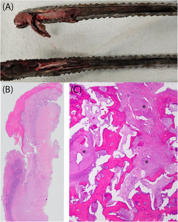
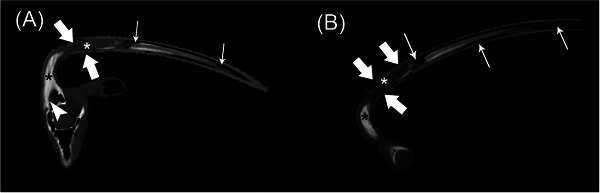
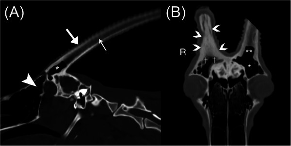
A 3-year-old male Scimitar-horned oryx presented for evaluation of an acutely deformed right horn with right head tilt and right facial pain. Computed tomographic evaluation revealed an increased volume of central fluid/soft tissue attenuation with gas-attenuating foci within the right horn. The right horn was amputated at the right horn base. Imaging and histopathologic features were consistent with emphysematous osteomyelitis. Following treatment, the patient returned to normal behavior. This is the first veterinary report describing the computed tomographic features of a horn infection in a Scimitar-horned oryx.
期刊介绍:
Veterinary Radiology & Ultrasound is a bimonthly, international, peer-reviewed, research journal devoted to the fields of veterinary diagnostic imaging and radiation oncology. Established in 1958, it is owned by the American College of Veterinary Radiology and is also the official journal for six affiliate veterinary organizations. Veterinary Radiology & Ultrasound is represented on the International Committee of Medical Journal Editors, World Association of Medical Editors, and Committee on Publication Ethics.
The mission of Veterinary Radiology & Ultrasound is to serve as a leading resource for high quality articles that advance scientific knowledge and standards of clinical practice in the areas of veterinary diagnostic radiology, computed tomography, magnetic resonance imaging, ultrasonography, nuclear imaging, radiation oncology, and interventional radiology. Manuscript types include original investigations, imaging diagnosis reports, review articles, editorials and letters to the Editor. Acceptance criteria include originality, significance, quality, reader interest, composition and adherence to author guidelines.


 分享
分享
 求助内容:
求助内容: 应助结果提醒方式:
 扫码关注我们
