Adi Porat Rein, Hashem Totah, Koby Brosh, David Zadok, Joel Hanhart
{"title":"Foveal hyper-reflective vertical lines detected by optical coherence tomography: Imaging features, literature review and differential diagnoses.","authors":"Adi Porat Rein, Hashem Totah, Koby Brosh, David Zadok, Joel Hanhart","doi":"10.1007/s00417-024-06616-5","DOIUrl":null,"url":null,"abstract":"<p><strong>Purpose: </strong>To describe foveal hyper-reflective vertical lines (FVL) as a specific morphological finding on structural spectral-domain optical coherence tomography (SD-OCT) and discuss its differential diagnosis.</p><p><strong>Methods: </strong>Observational case series. Ten patients (10 eyes) with FVL were meticulously examined at the Ophthalmology Department, Shaare Zedek Medical Center, Jerusalem, Israel. Detailed analysis of SD-OCT findings, clinical records, and retinal imaging was conducted to establish correlations between FVL and various underlying conditions.</p><p><strong>Results: </strong>We established the following list of settings, supported by the clinical context and ancillary investigations, in which SD-OCT displayed FVL: inflammation (1 eye), mechanical (1 eye), resorption of fluids of various origins (4 eyes), macular telangiectasia (1 eye), age-related macular degeneration (1 eye), diabetic retinopathy (1 eye) and scar (1 eye).</p><p><strong>Conclusions: </strong>FVL can be observed in various underlying conditions. Recognition of this pattern and formulation of an appropriate differential diagnosis is of interest for correctly diagnosing and treating patients whose structural OCT harbors this yet overlooked finding.</p>","PeriodicalId":12795,"journal":{"name":"Graefe’s Archive for Clinical and Experimental Ophthalmology","volume":" ","pages":"849-855"},"PeriodicalIF":2.4000,"publicationDate":"2025-03-01","publicationTypes":"Journal Article","fieldsOfStudy":null,"isOpenAccess":false,"openAccessPdf":"https://www.ncbi.nlm.nih.gov/pmc/articles/PMC11953122/pdf/","citationCount":"0","resultStr":null,"platform":"Semanticscholar","paperid":null,"PeriodicalName":"Graefe’s Archive for Clinical and Experimental Ophthalmology","FirstCategoryId":"3","ListUrlMain":"https://doi.org/10.1007/s00417-024-06616-5","RegionNum":3,"RegionCategory":"医学","ArticlePicture":[],"TitleCN":null,"AbstractTextCN":null,"PMCID":null,"EPubDate":"2024/10/15 0:00:00","PubModel":"Epub","JCR":"Q2","JCRName":"OPHTHALMOLOGY","Score":null,"Total":0}
引用次数: 0
Abstract
Purpose: To describe foveal hyper-reflective vertical lines (FVL) as a specific morphological finding on structural spectral-domain optical coherence tomography (SD-OCT) and discuss its differential diagnosis.
Methods: Observational case series. Ten patients (10 eyes) with FVL were meticulously examined at the Ophthalmology Department, Shaare Zedek Medical Center, Jerusalem, Israel. Detailed analysis of SD-OCT findings, clinical records, and retinal imaging was conducted to establish correlations between FVL and various underlying conditions.
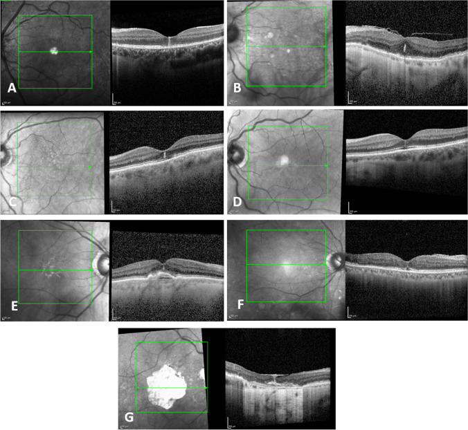
Results: We established the following list of settings, supported by the clinical context and ancillary investigations, in which SD-OCT displayed FVL: inflammation (1 eye), mechanical (1 eye), resorption of fluids of various origins (4 eyes), macular telangiectasia (1 eye), age-related macular degeneration (1 eye), diabetic retinopathy (1 eye) and scar (1 eye).
Conclusions: FVL can be observed in various underlying conditions. Recognition of this pattern and formulation of an appropriate differential diagnosis is of interest for correctly diagnosing and treating patients whose structural OCT harbors this yet overlooked finding.
期刊介绍:
Graefe''s Archive for Clinical and Experimental Ophthalmology is a distinguished international journal that presents original clinical reports and clini-cally relevant experimental studies. Founded in 1854 by Albrecht von Graefe to serve as a source of useful clinical information and a stimulus for discussion, the journal has published articles by leading ophthalmologists and vision research scientists for more than a century. With peer review by an international Editorial Board and prompt English-language publication, Graefe''s Archive provides rapid dissemination of clinical and clinically related experimental information.


 分享
分享
 求助内容:
求助内容: 应助结果提醒方式:
 扫码关注我们
