Marina Perez, Ainhoa Meseguer, Julio Vara, Jose Carlos Vilches, Ignacio Brunel, Manuel Lozano, Rodrigo Orozco, Juan Luis Alcazar
{"title":"GI-RADS versus O-RADS in the differential diagnosis of adnexal masses: a systematic review and head-to-head meta-analysis.","authors":"Marina Perez, Ainhoa Meseguer, Julio Vara, Jose Carlos Vilches, Ignacio Brunel, Manuel Lozano, Rodrigo Orozco, Juan Luis Alcazar","doi":"10.14366/usg.24105","DOIUrl":null,"url":null,"abstract":"<p><strong>Purpose: </strong>The aim of this study was to compare the diagnostic performance of the Gynecology Imaging Reporting and Data System (GI-RADS) and Ovarian-Adnexal Reporting and Data System (O-RADS) ultrasound (US) classification systems and assess their capacity to stratify the risk of malignancy in adnexal masses (AMs).</p><p><strong>Methods: </strong>A comprehensive search of MEDLINE (PubMed), Scopus, Web of Science, and Google Scholar was conducted to identify articles published between January 2020 and August 2023. The quality of the studies, the risk of bias, and concerns regarding applicability were assessed using QUADAS-2.</p><p><strong>Results: </strong>The search yielded 132 citations. Five articles, which included a total of 2,448 AMs, were ultimately selected for inclusion. The risk of bias was high in all articles regarding patient selection, low in four studies for the index test, and unclear in three papers for the reference test. For GI-RADS, the pooled sensitivity and specificity were 90.8% (95% confidence interval [CI], 86.0% to 94.0%) and 91.5% (95% CI, 89.0% to 93.0%), respectively. For O-RADS, the pooled sensitivity and specificity were 95.1% (95% CI, 93.0% to 97.0%) and 88.8% (95% CI, 85.0% to 92.0%), respectively. O-RADS demonstrated greater sensitivity for malignancy than GI-RADS (P<0.05). Heterogeneity was moderate for both sensitivity and specificity with respect to GIRADS; for O-RADS, heterogeneity was moderate for sensitivity and high for specificity.</p><p><strong>Conclusion: </strong>Both GI-RADS and O-RADS US demonstrate good diagnostic performance in the preoperative assessment of AMs. However, the O-RADS classification provides superior sensitivity.</p>","PeriodicalId":54227,"journal":{"name":"Ultrasonography","volume":" ","pages":"438-447"},"PeriodicalIF":2.5000,"publicationDate":"2024-11-01","publicationTypes":"Journal Article","fieldsOfStudy":null,"isOpenAccess":false,"openAccessPdf":"https://www.ncbi.nlm.nih.gov/pmc/articles/PMC11532524/pdf/","citationCount":"0","resultStr":null,"platform":"Semanticscholar","paperid":null,"PeriodicalName":"Ultrasonography","FirstCategoryId":"3","ListUrlMain":"https://doi.org/10.14366/usg.24105","RegionNum":3,"RegionCategory":"医学","ArticlePicture":[],"TitleCN":null,"AbstractTextCN":null,"PMCID":null,"EPubDate":"2024/9/2 0:00:00","PubModel":"Epub","JCR":"Q2","JCRName":"RADIOLOGY, NUCLEAR MEDICINE & MEDICAL IMAGING","Score":null,"Total":0}
引用次数: 0
Abstract
Purpose: The aim of this study was to compare the diagnostic performance of the Gynecology Imaging Reporting and Data System (GI-RADS) and Ovarian-Adnexal Reporting and Data System (O-RADS) ultrasound (US) classification systems and assess their capacity to stratify the risk of malignancy in adnexal masses (AMs).
Methods: A comprehensive search of MEDLINE (PubMed), Scopus, Web of Science, and Google Scholar was conducted to identify articles published between January 2020 and August 2023. The quality of the studies, the risk of bias, and concerns regarding applicability were assessed using QUADAS-2.
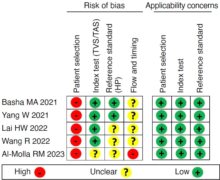
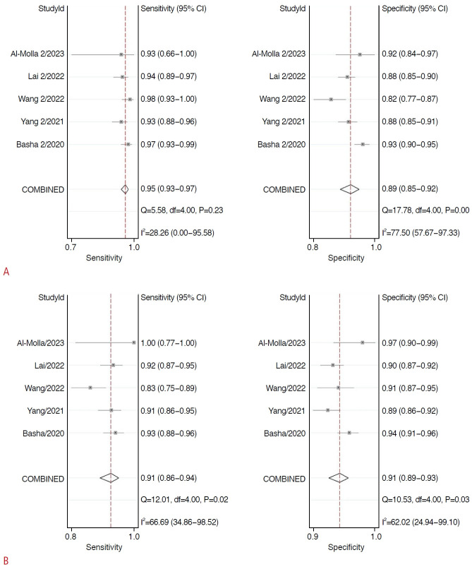
Results: The search yielded 132 citations. Five articles, which included a total of 2,448 AMs, were ultimately selected for inclusion. The risk of bias was high in all articles regarding patient selection, low in four studies for the index test, and unclear in three papers for the reference test. For GI-RADS, the pooled sensitivity and specificity were 90.8% (95% confidence interval [CI], 86.0% to 94.0%) and 91.5% (95% CI, 89.0% to 93.0%), respectively. For O-RADS, the pooled sensitivity and specificity were 95.1% (95% CI, 93.0% to 97.0%) and 88.8% (95% CI, 85.0% to 92.0%), respectively. O-RADS demonstrated greater sensitivity for malignancy than GI-RADS (P<0.05). Heterogeneity was moderate for both sensitivity and specificity with respect to GIRADS; for O-RADS, heterogeneity was moderate for sensitivity and high for specificity.
Conclusion: Both GI-RADS and O-RADS US demonstrate good diagnostic performance in the preoperative assessment of AMs. However, the O-RADS classification provides superior sensitivity.
UltrasonographyMedicine-Radiology, Nuclear Medicine and Imaging
CiteScore
5.10
自引率
6.50%
发文量
78
审稿时长
15 weeks
期刊介绍:
Ultrasonography, the official English-language journal of the Korean Society of Ultrasound in Medicine (KSUM), is an international peer-reviewed academic journal dedicated to practice, research, technology, and education dealing with medical ultrasound. It is renamed from the Journal of Korean Society of Ultrasound in Medicine in January 2014, and published four times per year: January 1, April 1, July 1, and October 1. Original articles, technical notes, topical reviews, perspectives, pictorial essays, and timely editorial materials are published in Ultrasonography covering state-of-the-art content.
Ultrasonography aims to provide updated information on new diagnostic concepts and technical developments, including experimental animal studies using new equipment in addition to well-designed reviews of contemporary issues in patient care. Along with running KSUM Open, the annual international congress of KSUM, Ultrasonography also serves as a medium for cooperation among physicians and specialists from around the world who are focusing on various ultrasound technology and disease problems and relevant basic science.


 分享
分享
 求助内容:
求助内容: 应助结果提醒方式:
 扫码关注我们
