Yu Qiao Zhang, Xiu Juan Zhang, Ru Yue Shen, Yuzhou Zhang, Fang Yao Tang, Simon K H Szeto, Danny Siu-Chun Ng, Ka Wai Kam, Alvin L Young, Li Jia Chen, Chi Pui Pang, Clement C Tham, Jason C Yam, Poemen P Chan
{"title":"Exploring optical coherence tomography parameters in eyes with myopic tilted disc.","authors":"Yu Qiao Zhang, Xiu Juan Zhang, Ru Yue Shen, Yuzhou Zhang, Fang Yao Tang, Simon K H Szeto, Danny Siu-Chun Ng, Ka Wai Kam, Alvin L Young, Li Jia Chen, Chi Pui Pang, Clement C Tham, Jason C Yam, Poemen P Chan","doi":"10.1186/s40662-024-00411-3","DOIUrl":null,"url":null,"abstract":"<p><strong>Background: </strong>To investigate the impact of optic disc torsion (ODT), horizontal disc tilt (HDT) angle, and ovality index (OI) on different retinal nerve fiber layer (RNFL) and ganglion cell-inner plexiform layer (GCIPL) segments in healthy myopic eyes.</p><p><strong>Methods: </strong>ODT and OI were measured from fundus photographs. HDT angle, peripapillary RNFL, and macular GCIPL were measured by swept-source optical coherence tomography (SS-OCT). The association between optic disc morphology and the RNFL/GCIPL thickness were evaluated, with age and axial length (AL) adjusted.</p><p><strong>Results: </strong>Among 530 healthy myopic eyes of 284 participants (mean age: 41.7 years, mean spherical equivalent: - 7.70 D, and mean AL: 26.6 mm), 335 eyes (63.2%) had temporal disc torsion (temporal group) and 195 eyes (36.8%) had nasal disc torsion (nasal group). For the nasal group, a larger OI was associated with thinner superior-to-superonasal GCIPL (β = - 7.465 to - 6.972, both P = 0.024) and temporal RNFL sectors (β = - 49.596 to - 27.748, P ≤ 0.014). For the temporal group, a larger OI was associated with thinner superior-to-nasal (β = - 50.255 to - 22.093, P ≤ 0.006) and thicker temporal RNFL sectors (β = 29.015 to 56.890, P ≤ 0.003). Additionally, a larger HDT angle was associated with thinner superior-to-nasal RNFL sectors (β = - 0.559 to - 0.242, P ≤ 0.036) and thinner superior-to-superotemporal GCIPL sectors (β = - 0.084 to - 0.069, P ≤ 0.037).</p><p><strong>Conclusions: </strong>The optic disc tortional direction was associated with the measurement of different RNFL and GCIPL sectors independent of the AL and age. These should be considered when constructing a myopic normative database.</p>","PeriodicalId":12194,"journal":{"name":"Eye and Vision","volume":"11 1","pages":"47"},"PeriodicalIF":4.0000,"publicationDate":"2024-11-02","publicationTypes":"Journal Article","fieldsOfStudy":null,"isOpenAccess":false,"openAccessPdf":"https://www.ncbi.nlm.nih.gov/pmc/articles/PMC11580533/pdf/","citationCount":"0","resultStr":null,"platform":"Semanticscholar","paperid":null,"PeriodicalName":"Eye and Vision","FirstCategoryId":"3","ListUrlMain":"https://doi.org/10.1186/s40662-024-00411-3","RegionNum":1,"RegionCategory":"医学","ArticlePicture":[],"TitleCN":null,"AbstractTextCN":null,"PMCID":null,"EPubDate":"","PubModel":"","JCR":"Q1","JCRName":"OPHTHALMOLOGY","Score":null,"Total":0}
引用次数: 0
Abstract
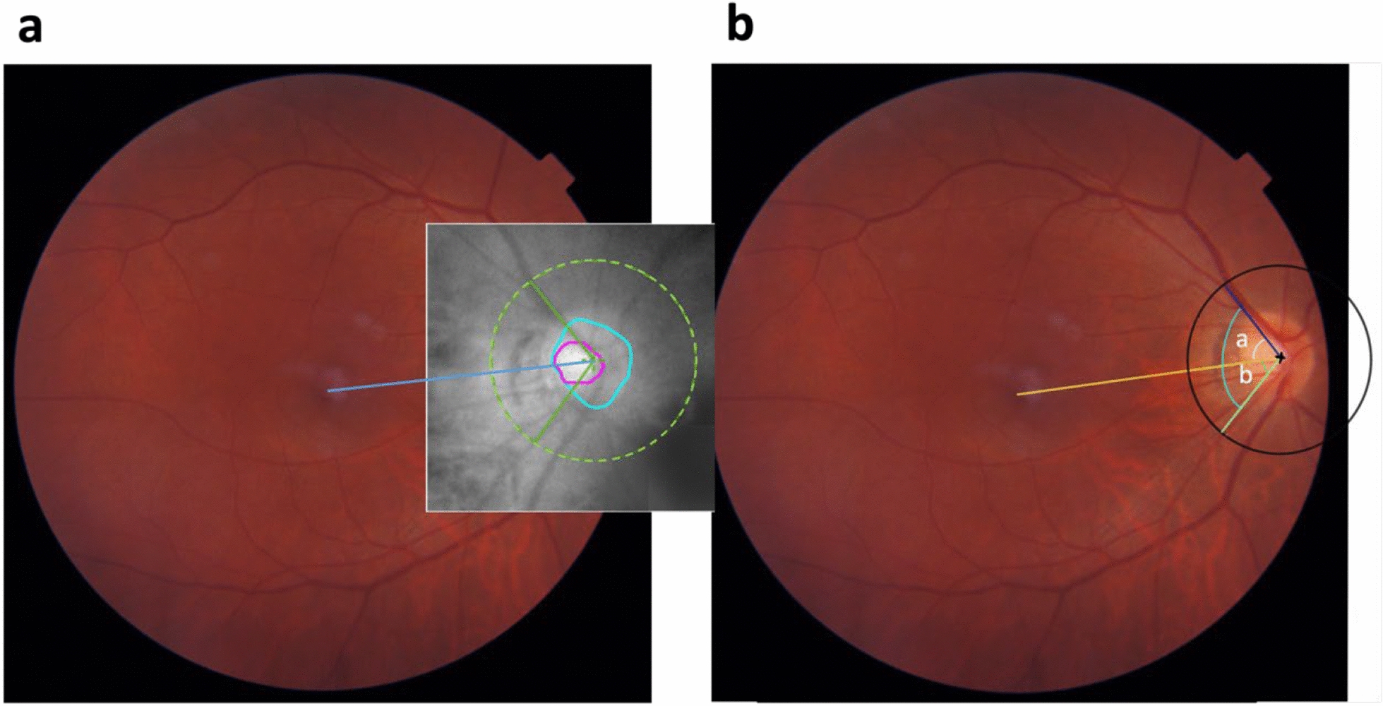
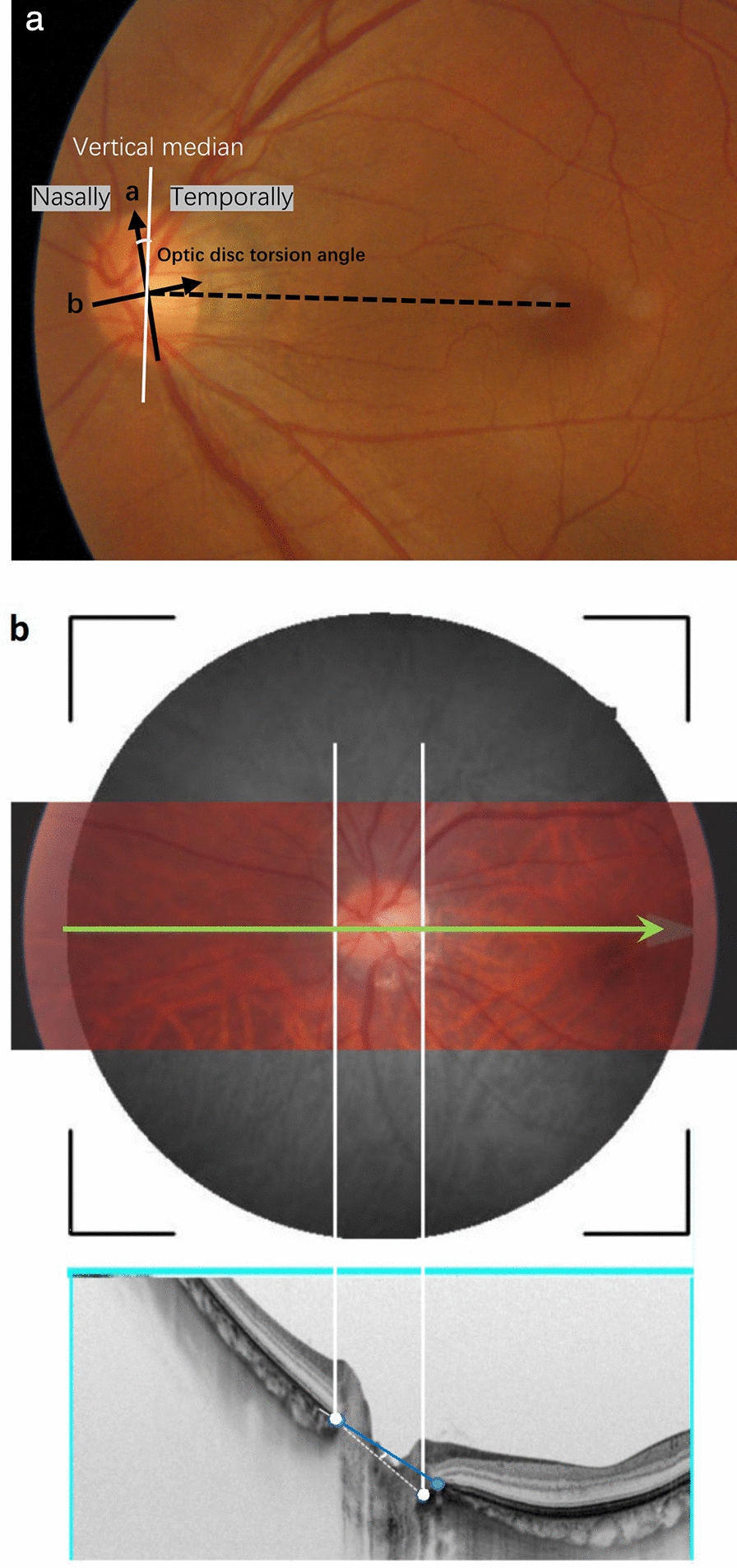
Background: To investigate the impact of optic disc torsion (ODT), horizontal disc tilt (HDT) angle, and ovality index (OI) on different retinal nerve fiber layer (RNFL) and ganglion cell-inner plexiform layer (GCIPL) segments in healthy myopic eyes.
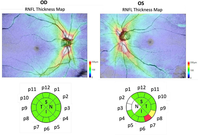
Methods: ODT and OI were measured from fundus photographs. HDT angle, peripapillary RNFL, and macular GCIPL were measured by swept-source optical coherence tomography (SS-OCT). The association between optic disc morphology and the RNFL/GCIPL thickness were evaluated, with age and axial length (AL) adjusted.
Results: Among 530 healthy myopic eyes of 284 participants (mean age: 41.7 years, mean spherical equivalent: - 7.70 D, and mean AL: 26.6 mm), 335 eyes (63.2%) had temporal disc torsion (temporal group) and 195 eyes (36.8%) had nasal disc torsion (nasal group). For the nasal group, a larger OI was associated with thinner superior-to-superonasal GCIPL (β = - 7.465 to - 6.972, both P = 0.024) and temporal RNFL sectors (β = - 49.596 to - 27.748, P ≤ 0.014). For the temporal group, a larger OI was associated with thinner superior-to-nasal (β = - 50.255 to - 22.093, P ≤ 0.006) and thicker temporal RNFL sectors (β = 29.015 to 56.890, P ≤ 0.003). Additionally, a larger HDT angle was associated with thinner superior-to-nasal RNFL sectors (β = - 0.559 to - 0.242, P ≤ 0.036) and thinner superior-to-superotemporal GCIPL sectors (β = - 0.084 to - 0.069, P ≤ 0.037).
Conclusions: The optic disc tortional direction was associated with the measurement of different RNFL and GCIPL sectors independent of the AL and age. These should be considered when constructing a myopic normative database.
期刊介绍:
Eye and Vision is an open access, peer-reviewed journal for ophthalmologists and visual science specialists. It welcomes research articles, reviews, methodologies, commentaries, case reports, perspectives and short reports encompassing all aspects of eye and vision. Topics of interest include but are not limited to: current developments of theoretical, experimental and clinical investigations in ophthalmology, optometry and vision science which focus on novel and high-impact findings on central issues pertaining to biology, pathophysiology and etiology of eye diseases as well as advances in diagnostic techniques, surgical treatment, instrument updates, the latest drug findings, results of clinical trials and research findings. It aims to provide ophthalmologists and visual science specialists with the latest developments in theoretical, experimental and clinical investigations in eye and vision.


 分享
分享
 求助内容:
求助内容: 应助结果提醒方式:
 扫码关注我们
