Xuewei Xie, Qianmei Jiang, Yue Suo, Chong Han, Zhaobin Wang, Zhe Zhang, Ning Wang, Yihuai Wang, Chunguang Zhang, Bingshan Xue, Tao Liu, David Wang, Jing Jing, Yongjun Wang
{"title":"0.23-Tesla MRI to differentiate between ischaemic and haemorrhagic strokes within 24 hours of onset: a combined experimental-clinical study.","authors":"Xuewei Xie, Qianmei Jiang, Yue Suo, Chong Han, Zhaobin Wang, Zhe Zhang, Ning Wang, Yihuai Wang, Chunguang Zhang, Bingshan Xue, Tao Liu, David Wang, Jing Jing, Yongjun Wang","doi":"10.1136/svn-2024-003592","DOIUrl":null,"url":null,"abstract":"<p><strong>Background and purpose: </strong>The low-field MRI is a promising tool to accurately diagnose strokes. We here report our study on the accuracy of a 0.23-Tesla (0.23-T) MRI using the haematoma enhanced inversion recovery (HEIR) sequence to detect acute ischaemic stroke (AIS) and intracerebral haemorrhage (ICH) within 24 hours of symptom onset.</p><p><strong>Methods: </strong>A novel HEIR sequence based on fluid-attenuated inversion recovery T1-weighted, with a scanning time of 1 min and 17 s, was developed using an ICH and AIS pig model on a 0.23-T MRI. Images of the pig model were obtained hourly for 24 hours in order to monitor value changes on T1/T2 and verify the differential diagnosis of AIS and ICH. Then, 30 patients with AIS and 30 patients with ICH with confirmed diagnoses by 3T-MRI/CT were included. Diagnostic criteria on a 0.23-T MRI for ICH was the hyperintensity signal on both the diffusion-weighted imaging (DWI) and HEIR sequence, while for AIS was the hyperintensity on DWI and isointensity on the HEIR sequence. Two blinded raters independently assessed the images obtained by the 0.23-T MRI for the presence of ICH/AIS.</p><p><strong>Results: </strong>In the pig model, setting the inversion time to 800 ms enabled clear differentiation of ICH from brain parenchymal tissue and AIS. In real patients, a correct 0.23-T MRI diagnosis of either an AIS or ICH was made in all 60 patients within 24 hours of symptom onset (100% overall accuracy). No adverse events occurred.</p><p><strong>Conclusions: </strong>The 0.23-T MRI may have the potential to differentiate cerebral haemorrhage from cerebral infarction with both speed and accuracy, making brain MRI scans easier, faster and cheaper. It might be possible to improve the screening imaging process for strokes in the emergency room. Further multicentre studies are needed to validate our findings.</p>","PeriodicalId":48733,"journal":{"name":"Journal of Investigative Medicine","volume":" ","pages":"472-480"},"PeriodicalIF":4.9000,"publicationDate":"2025-08-26","publicationTypes":"Journal Article","fieldsOfStudy":null,"isOpenAccess":false,"openAccessPdf":"https://www.ncbi.nlm.nih.gov/pmc/articles/PMC12415649/pdf/","citationCount":"0","resultStr":null,"platform":"Semanticscholar","paperid":null,"PeriodicalName":"Journal of Investigative Medicine","FirstCategoryId":"3","ListUrlMain":"https://doi.org/10.1136/svn-2024-003592","RegionNum":1,"RegionCategory":"医学","ArticlePicture":[],"TitleCN":null,"AbstractTextCN":null,"PMCID":null,"EPubDate":"","PubModel":"","JCR":"","JCRName":"","Score":null,"Total":0}
引用次数: 0
Abstract
Background and purpose: The low-field MRI is a promising tool to accurately diagnose strokes. We here report our study on the accuracy of a 0.23-Tesla (0.23-T) MRI using the haematoma enhanced inversion recovery (HEIR) sequence to detect acute ischaemic stroke (AIS) and intracerebral haemorrhage (ICH) within 24 hours of symptom onset.
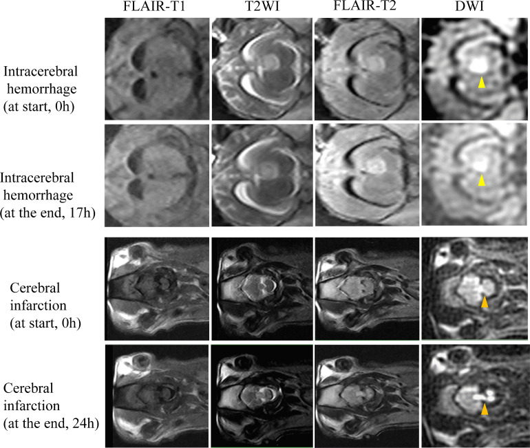
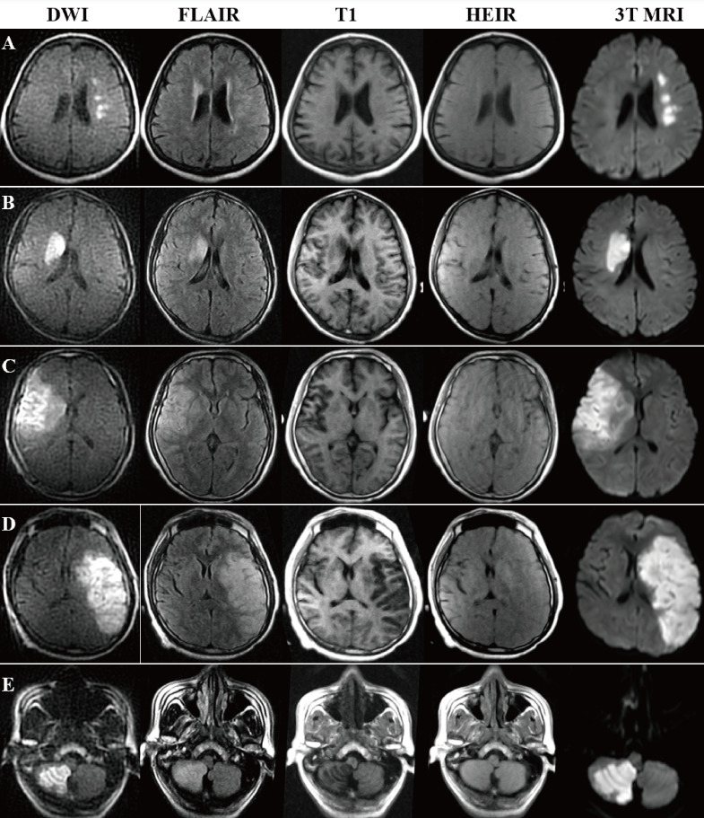
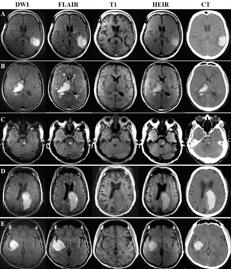
Methods: A novel HEIR sequence based on fluid-attenuated inversion recovery T1-weighted, with a scanning time of 1 min and 17 s, was developed using an ICH and AIS pig model on a 0.23-T MRI. Images of the pig model were obtained hourly for 24 hours in order to monitor value changes on T1/T2 and verify the differential diagnosis of AIS and ICH. Then, 30 patients with AIS and 30 patients with ICH with confirmed diagnoses by 3T-MRI/CT were included. Diagnostic criteria on a 0.23-T MRI for ICH was the hyperintensity signal on both the diffusion-weighted imaging (DWI) and HEIR sequence, while for AIS was the hyperintensity on DWI and isointensity on the HEIR sequence. Two blinded raters independently assessed the images obtained by the 0.23-T MRI for the presence of ICH/AIS.
Results: In the pig model, setting the inversion time to 800 ms enabled clear differentiation of ICH from brain parenchymal tissue and AIS. In real patients, a correct 0.23-T MRI diagnosis of either an AIS or ICH was made in all 60 patients within 24 hours of symptom onset (100% overall accuracy). No adverse events occurred.
Conclusions: The 0.23-T MRI may have the potential to differentiate cerebral haemorrhage from cerebral infarction with both speed and accuracy, making brain MRI scans easier, faster and cheaper. It might be possible to improve the screening imaging process for strokes in the emergency room. Further multicentre studies are needed to validate our findings.
期刊介绍:
Journal of Investigative Medicine (JIM) is the official publication of the American Federation for Medical Research. The journal is peer-reviewed and publishes high-quality original articles and reviews in the areas of basic, clinical, and translational medical research.
JIM publishes on all topics and specialty areas that are critical to the conduct of the entire spectrum of biomedical research: from the translation of clinical observations at the bedside, to basic and animal research to clinical research and the implementation of innovative medical care.


 分享
分享
 求助内容:
求助内容: 应助结果提醒方式:
 扫码关注我们
