Marisol De La Rosa-Alamilla, Rubí Chávez-Silva, Herminia Uscanga-Carrasco
{"title":"[Use of ultrasound in vein of Galen malformation].","authors":"Marisol De La Rosa-Alamilla, Rubí Chávez-Silva, Herminia Uscanga-Carrasco","doi":"10.5281/zenodo.13306799","DOIUrl":null,"url":null,"abstract":"<p><strong>Background: </strong>Aneurysmal malformation of the vein of Galen (AVMG) is a rare malformation of the intracranial venous system. The incidence is estimated at 1:10,000-25,0000 births. The clinical picture is very variable, in the neonatal period it includes seizures and heart failure. It is very difficult to diagnose, but by doing it in a timely manner, mortality is greatly reduced. It can be detected by prenatal ultrasounds and, at birth, the gold standard imaging study for its diagnosis is cerebral resonance and angioresonance. However, 50% of AVMG cannot be corrected and there is evidence that 77% of untreated cases result in death.</p><p><strong>Clinical case: </strong>The case of a newborn with a prenatal diagnosis is presented and at birth the diagnosis of MAVG was corroborated by neurosonography, and subsequently confirmed by venoresonance. His clinical evolution was documented, which included placement of a ventriculoperitoneal shunt and vascular embolization; however, there was a fatal outcome.</p><p><strong>Conclusions: </strong>In MAVG it is important to define the anatomy of the lesion, due to the clinical, therapeutic and prognostic implications that this represents. B-mode ultrasound, together with Doppler mode, are essential complementary tools for the diagnosis of AVMG, as well as for prognostic evaluation and to provide information for counseling parents and for optimal management. Successful treatment remains a complex therapeutic challenge.</p>","PeriodicalId":94200,"journal":{"name":"Revista medica del Instituto Mexicano del Seguro Social","volume":"62 6","pages":"1-5"},"PeriodicalIF":0.0000,"publicationDate":"2024-11-04","publicationTypes":"Journal Article","fieldsOfStudy":null,"isOpenAccess":false,"openAccessPdf":"https://www.ncbi.nlm.nih.gov/pmc/articles/PMC12549054/pdf/","citationCount":"0","resultStr":null,"platform":"Semanticscholar","paperid":null,"PeriodicalName":"Revista medica del Instituto Mexicano del Seguro Social","FirstCategoryId":"1085","ListUrlMain":"https://doi.org/10.5281/zenodo.13306799","RegionNum":0,"RegionCategory":null,"ArticlePicture":[],"TitleCN":null,"AbstractTextCN":null,"PMCID":null,"EPubDate":"","PubModel":"","JCR":"","JCRName":"","Score":null,"Total":0}
引用次数: 0
Abstract
Background: Aneurysmal malformation of the vein of Galen (AVMG) is a rare malformation of the intracranial venous system. The incidence is estimated at 1:10,000-25,0000 births. The clinical picture is very variable, in the neonatal period it includes seizures and heart failure. It is very difficult to diagnose, but by doing it in a timely manner, mortality is greatly reduced. It can be detected by prenatal ultrasounds and, at birth, the gold standard imaging study for its diagnosis is cerebral resonance and angioresonance. However, 50% of AVMG cannot be corrected and there is evidence that 77% of untreated cases result in death.
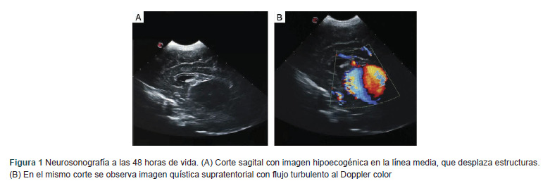
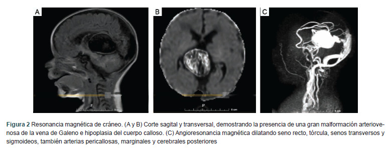
Clinical case: The case of a newborn with a prenatal diagnosis is presented and at birth the diagnosis of MAVG was corroborated by neurosonography, and subsequently confirmed by venoresonance. His clinical evolution was documented, which included placement of a ventriculoperitoneal shunt and vascular embolization; however, there was a fatal outcome.
Conclusions: In MAVG it is important to define the anatomy of the lesion, due to the clinical, therapeutic and prognostic implications that this represents. B-mode ultrasound, together with Doppler mode, are essential complementary tools for the diagnosis of AVMG, as well as for prognostic evaluation and to provide information for counseling parents and for optimal management. Successful treatment remains a complex therapeutic challenge.


 分享
分享

 求助内容:
求助内容: 应助结果提醒方式:
 扫码关注我们
