{"title":"Targeting LAG-3 restores CD8+ T cell effector function through CD94/NKG2-Qa-1b signaling","authors":"Zhiqiang Wang, Mingzhu Yin, Ge Lou","doi":"10.1002/mog2.70003","DOIUrl":null,"url":null,"abstract":"<p>A recent study published in <i>Cell</i> by Ngiow et al. elucidates the synergistic roles of programmed cell death protein 1 (PD-1) and lymphocyte-activation gene 3 (LAG-3) in CD8<sup>+</sup> T cells.<span><sup>1</sup></span> The authors demonstrate that LAG-3 influences the fate of these cells by sustaining thymocyte selection-associated high mobility group box protein (TOX) expression and modulating the CD94/NKG2-Qa-1b axis in exhausted CD8<sup>+</sup> T cells. This research enhances our understanding of the intricate mechanisms underlying immune checkpoint blockade and paves the way for novel approaches in cancer immunotherapy.</p><p>In chronic viral infections and cancers, CD8<sup>+</sup> T cells, known as the “warriors” of the immune system, frequently experience exhaustion due to persistent antigenic stimulation. This exhaustion results in impaired proliferation and effector function of exhausted CD8<sup>+</sup> T cells (Tex), ultimately leading to immune failure. The defining characteristics of Tex include a distinct TOX-driven transcriptional and epigenetic state, along with the sustained expression of various inhibitory receptors (IRs), such as PD-1, LAG-3, and cytotoxic T-lymphocyte antigen-4 (CTLA-4).<span><sup>2</sup></span> Recent advancements in immune checkpoint blockade therapies that target various IRs have significantly improved tumor treatment outcomes. However, only a subset of tumor patients exhibit sustained clinical responses to monotherapy with PD-1/programmed death-ligand 1 (PD-L1) blockade. Consequently, there is a growing interest in exploring the combination of multiple immune receptors to enhance the efficacy of immunotherapeutic approaches. A recent phase III clinical trial, RELATIVITY-047, demonstrated that combining a LAG-3 antibody (relatlimab) and a PD-1 antibody (nivolumab) exhibits enhanced clinical antitumor effects. However, the precise mechanisms by which PD-1 and LAG-3 regulate Tex function, as well as the interconnections between their signaling pathways, remain unclear.</p><p>Ngiow et al. investigated the effects of LAG-3 and PD-1 on CD8<sup>+</sup> T cells at various stages of chronic infection using the Quad transplantation model. Their findings revealed that CD8<sup>+</sup> T cells lacking PD-1 proliferated significantly faster during the early stages of infection; however, these cells became progressively less sustainable over time. In contrast, cells lacking LAG-3 demonstrated enhanced effector functions, particularly in terms of cytotoxicity and cytokine secretion. This indicates that PD-1 primarily inhibits cell proliferation, while LAG-3 restricts the multifunctionality and killing capacity of effector T cells as they transition into exhausted T cells. The researchers further investigated the mechanism by which LAG-3 influences the differentiation of Tex cells through the regulation of TOX expression. Their findings revealed that LAG-3 deficiency resulted in a significant reduction in TOX expression, which was accompanied by a decrease in TCF1<sup>+</sup> Tex precursor cells. This suggests that LAG-3 plays a crucial role in sustaining the persistence and functionality of Tex cells by maintaining TOX levels.</p><p>Additionally, RNA sequencing results indicate that LAG-3-deficient cells exhibit a transcriptional profile more closely aligned with effector T cells, showing enhanced expression of cytotoxicity-associated genes. These genes, which include multiple members of the Klr family (e.g., Klrg1 and Klrd1), indicate that LAG-3 is crucial in suppressing the expression of natural killer (NK) receptor-related genes. Bioinformatic data analysis further revealed that LAG-3 knockout (KO) cells exhibited an enrichment in pathways related to NK cell-mediated cytotoxicity. These findings not only underscore the significance of LAG-3 in the regulation of Tex cell function and differentiation but also suggest that LAG-3 may influence cell-killing abilities and immune surveillance through its regulation of NK receptors. The researchers subsequently investigated the transcriptomic profiles of PD-1 KO and LAG-3 KO cells using single-cell RNA sequencing, comparing them with TOX-deficient cells. The results indicated that LAG-3-deficient Tex cells displayed elevated expression of genes associated with NK receptors, such as CD94 and Klrd1.</p><p>The experimental results indicated an upregulation of CD94 expression in LAG-3-deficient cells, which was associated with enhanced NK cell-like functionality. This enhancement was particularly pronounced within the tumor microenvironment, suggesting that a key mechanism by which LAG-3 regulates Tex is through the inhibition of CD94/NKG2 axis activation. The investigators observed that LAG-3-deficient Tex cells exhibited a higher expression of the activating receptor NK cell Group 2 isoform C/E (NKG2C/E) compared to the inhibitory receptor NK cell lectin-like receptor subfamily C member 1 (NKG2A). This shift in expression patterns enabled these cells to more effectively target and eliminate tumor cells within the microenvironment.</p><p>Further experiments demonstrated that LAG-3-deficient Tex cells displayed an enhanced tumor-killing capacity that is dependent on the Qa-1b molecule. Qa-1b is a nonclassical MHC molecule expressed by tumor cells in response to stress, and tumor cells often evade immune surveillance by upregulating Qa-1b expression. By regulating the function of CD94/NKG2A, novel immunotherapeutic strategies can be designed to enhance the recognition and killing of tumor cells by T cells. The deletion of LAG-3 resulted in the upregulation of NKG2C/E-activated receptors in Tex cells, thereby transforming the expression of Qa-1b from a mechanism of tumor escape to one that is sensitive to tumors (Figure 1). This finding suggests that the inhibition of LAG-3 and PD-1 in tumor immunotherapy may enhance antitumor responses by modifying the expression profile of NK receptors. Consequently, this study not only elucidates the role of LAG-3 in immune checkpoint blockade but also highlights the potential for combining LAG-3 and PD-1, particularly in the context of Qa-1b-related tumor immunosurveillance. NKG2A blockade may be beneficial in combination with PD-1 blockade but may have limited effect in combination with anti-LAG-3/PD-1. This is because the primary effect of LAG-3 blockade is to deregulate inhibitory signaling in NK cells, and further inhibition of NKG2A may result in diminishing benefits.<span><sup>3</sup></span></p><p>The functions of PD-1 and LAG-3 in T-cell depletion are not merely superimposed; rather, they demonstrate nonredundant and unique synergistic effects. A deeper understanding of these nonredundant mechanisms not only enhances our comprehension of the immune system but also offers novel insights into the optimization of PD-1 and LAG-3 blockade strategies in future immunotherapy. The research conducted by Andrews and Cillo et al. demonstrated the coordinated effects of PD-1 and LAG-3 in promoting exhausted CD8<sup>+</sup> T cells, drawing on both clinical data and laboratory analysis.<span><sup>4, 5</sup></span> Their findings provide valuable insights into the combined effects of inhibitory receptors, establishing a theoretical basis and offering research examples that could inform clinical strategies for anti-infection and antitumor treatments.</p><p>This study has several limitations worth noting. First, the research primarily focuses on mouse models of chronic LCMV infection and extends to murine tumor models and human cancer patients, which may limit the generalizability of findings due to potential context-specific interactions or dependencies on particular LAG-3 ligands. Second, while the Quad transfer system effectively controls intrinsic cellular effects and accounts for variations in antigen load and inflammation, further investigation into antibody-mediated blockade, including its impact on LAG-3<sup>+</sup> cells beyond T ex cells, is necessary. Thirdly, although the study emphasizes CD94/NKG2 receptors, a more comprehensive analysis of other NK receptors like NKG2D is essential for a complete understanding of NK cell function. Finally, despite the promising prospects of LAG-3 as a novel immunomodulatory target for both cancer immunotherapy and infectious disease treatment, future studies must address challenges such as safety concerns, biomarker identification, and the complexities of the tumor microenvironment to translate these insights into effective clinical applications.</p><p><b>Zhiqiang Wang</b> and <b>Mingzhu Yin</b>: Conceptualization (equal); supervision (equal); writing—review and editing (equal). <b>Zhiqiang Wang</b> and <b>Ge Lou</b>: Conceptualization (equal); methodology (equal); project administration (equal); supervision (equal). All authors have read and approved the final manuscript.</p><p>The authors declare no conflict of interest.</p><p>The ethics statement is not available.</p>","PeriodicalId":100902,"journal":{"name":"MedComm – Oncology","volume":"3 4","pages":""},"PeriodicalIF":2.2000,"publicationDate":"2024-11-24","publicationTypes":"Journal Article","fieldsOfStudy":null,"isOpenAccess":false,"openAccessPdf":"https://onlinelibrary.wiley.com/doi/epdf/10.1002/mog2.70003","citationCount":"0","resultStr":null,"platform":"Semanticscholar","paperid":null,"PeriodicalName":"MedComm – Oncology","FirstCategoryId":"1085","ListUrlMain":"https://onlinelibrary.wiley.com/doi/10.1002/mog2.70003","RegionNum":0,"RegionCategory":null,"ArticlePicture":[],"TitleCN":null,"AbstractTextCN":null,"PMCID":null,"EPubDate":"","PubModel":"","JCR":"","JCRName":"","Score":null,"Total":0}
引用次数: 0
Abstract
A recent study published in Cell by Ngiow et al. elucidates the synergistic roles of programmed cell death protein 1 (PD-1) and lymphocyte-activation gene 3 (LAG-3) in CD8+ T cells.1 The authors demonstrate that LAG-3 influences the fate of these cells by sustaining thymocyte selection-associated high mobility group box protein (TOX) expression and modulating the CD94/NKG2-Qa-1b axis in exhausted CD8+ T cells. This research enhances our understanding of the intricate mechanisms underlying immune checkpoint blockade and paves the way for novel approaches in cancer immunotherapy.
In chronic viral infections and cancers, CD8+ T cells, known as the “warriors” of the immune system, frequently experience exhaustion due to persistent antigenic stimulation. This exhaustion results in impaired proliferation and effector function of exhausted CD8+ T cells (Tex), ultimately leading to immune failure. The defining characteristics of Tex include a distinct TOX-driven transcriptional and epigenetic state, along with the sustained expression of various inhibitory receptors (IRs), such as PD-1, LAG-3, and cytotoxic T-lymphocyte antigen-4 (CTLA-4).2 Recent advancements in immune checkpoint blockade therapies that target various IRs have significantly improved tumor treatment outcomes. However, only a subset of tumor patients exhibit sustained clinical responses to monotherapy with PD-1/programmed death-ligand 1 (PD-L1) blockade. Consequently, there is a growing interest in exploring the combination of multiple immune receptors to enhance the efficacy of immunotherapeutic approaches. A recent phase III clinical trial, RELATIVITY-047, demonstrated that combining a LAG-3 antibody (relatlimab) and a PD-1 antibody (nivolumab) exhibits enhanced clinical antitumor effects. However, the precise mechanisms by which PD-1 and LAG-3 regulate Tex function, as well as the interconnections between their signaling pathways, remain unclear.
Ngiow et al. investigated the effects of LAG-3 and PD-1 on CD8+ T cells at various stages of chronic infection using the Quad transplantation model. Their findings revealed that CD8+ T cells lacking PD-1 proliferated significantly faster during the early stages of infection; however, these cells became progressively less sustainable over time. In contrast, cells lacking LAG-3 demonstrated enhanced effector functions, particularly in terms of cytotoxicity and cytokine secretion. This indicates that PD-1 primarily inhibits cell proliferation, while LAG-3 restricts the multifunctionality and killing capacity of effector T cells as they transition into exhausted T cells. The researchers further investigated the mechanism by which LAG-3 influences the differentiation of Tex cells through the regulation of TOX expression. Their findings revealed that LAG-3 deficiency resulted in a significant reduction in TOX expression, which was accompanied by a decrease in TCF1+ Tex precursor cells. This suggests that LAG-3 plays a crucial role in sustaining the persistence and functionality of Tex cells by maintaining TOX levels.
Additionally, RNA sequencing results indicate that LAG-3-deficient cells exhibit a transcriptional profile more closely aligned with effector T cells, showing enhanced expression of cytotoxicity-associated genes. These genes, which include multiple members of the Klr family (e.g., Klrg1 and Klrd1), indicate that LAG-3 is crucial in suppressing the expression of natural killer (NK) receptor-related genes. Bioinformatic data analysis further revealed that LAG-3 knockout (KO) cells exhibited an enrichment in pathways related to NK cell-mediated cytotoxicity. These findings not only underscore the significance of LAG-3 in the regulation of Tex cell function and differentiation but also suggest that LAG-3 may influence cell-killing abilities and immune surveillance through its regulation of NK receptors. The researchers subsequently investigated the transcriptomic profiles of PD-1 KO and LAG-3 KO cells using single-cell RNA sequencing, comparing them with TOX-deficient cells. The results indicated that LAG-3-deficient Tex cells displayed elevated expression of genes associated with NK receptors, such as CD94 and Klrd1.
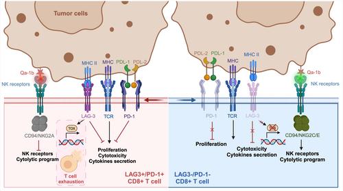
The experimental results indicated an upregulation of CD94 expression in LAG-3-deficient cells, which was associated with enhanced NK cell-like functionality. This enhancement was particularly pronounced within the tumor microenvironment, suggesting that a key mechanism by which LAG-3 regulates Tex is through the inhibition of CD94/NKG2 axis activation. The investigators observed that LAG-3-deficient Tex cells exhibited a higher expression of the activating receptor NK cell Group 2 isoform C/E (NKG2C/E) compared to the inhibitory receptor NK cell lectin-like receptor subfamily C member 1 (NKG2A). This shift in expression patterns enabled these cells to more effectively target and eliminate tumor cells within the microenvironment.
Further experiments demonstrated that LAG-3-deficient Tex cells displayed an enhanced tumor-killing capacity that is dependent on the Qa-1b molecule. Qa-1b is a nonclassical MHC molecule expressed by tumor cells in response to stress, and tumor cells often evade immune surveillance by upregulating Qa-1b expression. By regulating the function of CD94/NKG2A, novel immunotherapeutic strategies can be designed to enhance the recognition and killing of tumor cells by T cells. The deletion of LAG-3 resulted in the upregulation of NKG2C/E-activated receptors in Tex cells, thereby transforming the expression of Qa-1b from a mechanism of tumor escape to one that is sensitive to tumors (Figure 1). This finding suggests that the inhibition of LAG-3 and PD-1 in tumor immunotherapy may enhance antitumor responses by modifying the expression profile of NK receptors. Consequently, this study not only elucidates the role of LAG-3 in immune checkpoint blockade but also highlights the potential for combining LAG-3 and PD-1, particularly in the context of Qa-1b-related tumor immunosurveillance. NKG2A blockade may be beneficial in combination with PD-1 blockade but may have limited effect in combination with anti-LAG-3/PD-1. This is because the primary effect of LAG-3 blockade is to deregulate inhibitory signaling in NK cells, and further inhibition of NKG2A may result in diminishing benefits.3
The functions of PD-1 and LAG-3 in T-cell depletion are not merely superimposed; rather, they demonstrate nonredundant and unique synergistic effects. A deeper understanding of these nonredundant mechanisms not only enhances our comprehension of the immune system but also offers novel insights into the optimization of PD-1 and LAG-3 blockade strategies in future immunotherapy. The research conducted by Andrews and Cillo et al. demonstrated the coordinated effects of PD-1 and LAG-3 in promoting exhausted CD8+ T cells, drawing on both clinical data and laboratory analysis.4, 5 Their findings provide valuable insights into the combined effects of inhibitory receptors, establishing a theoretical basis and offering research examples that could inform clinical strategies for anti-infection and antitumor treatments.
This study has several limitations worth noting. First, the research primarily focuses on mouse models of chronic LCMV infection and extends to murine tumor models and human cancer patients, which may limit the generalizability of findings due to potential context-specific interactions or dependencies on particular LAG-3 ligands. Second, while the Quad transfer system effectively controls intrinsic cellular effects and accounts for variations in antigen load and inflammation, further investigation into antibody-mediated blockade, including its impact on LAG-3+ cells beyond T ex cells, is necessary. Thirdly, although the study emphasizes CD94/NKG2 receptors, a more comprehensive analysis of other NK receptors like NKG2D is essential for a complete understanding of NK cell function. Finally, despite the promising prospects of LAG-3 as a novel immunomodulatory target for both cancer immunotherapy and infectious disease treatment, future studies must address challenges such as safety concerns, biomarker identification, and the complexities of the tumor microenvironment to translate these insights into effective clinical applications.
Zhiqiang Wang and Mingzhu Yin: Conceptualization (equal); supervision (equal); writing—review and editing (equal). Zhiqiang Wang and Ge Lou: Conceptualization (equal); methodology (equal); project administration (equal); supervision (equal). All authors have read and approved the final manuscript.


 分享
分享

 求助内容:
求助内容: 应助结果提醒方式:
 扫码关注我们
