{"title":"Micro-CT Microcalcification Analysis: A Scoping Review of Current Applications and Future Potential in Breast Cancer Research.","authors":"Redona Brahimetaj, Jan Cornelis, Bart Jansen","doi":"10.3390/tomography10110126","DOIUrl":null,"url":null,"abstract":"<p><p>Micro-computed tomography (micro-CT) is a non-destructive imaging technique that offers highly detailed, 3D visualizations of a target specimen. In the context of breast cancer, micro-CT has emerged as a promising tool for analyzing microcalcifications (MCs), tiny calcium deposits that can indicate at an early stage the presence of cancer. This review aimed to explore the current applications of micro-CT in analyzing breast MCs (ex vivo, animal models, and phantoms) and to identify potential avenues in scientific research. We followed PRISMA guidelines for scoping reviews, yielding 18 studies that met our criteria. The studies varied in their purposes: feasibility and optimization of micro-CT for breast cancer imaging and MC analysis/diagnosis, comparison with other imaging modalities, development of micro-CT scanners and processing algorithms, enhancement of MC detection through contrast agents, etc. In conclusion, micro-CT offers superior image quality and detailed visualization of breast tissue (especially tumor masses and MCs), surpassing traditional methods like mammography and approaching the level of detail of histology. It holds great potential to enhance our understanding of MC characteristics and breast pathologies when used as a supplementary tool. Further research will solidify its role in clinical practice and potentially expand its applications in breast cancer studies.</p>","PeriodicalId":51330,"journal":{"name":"Tomography","volume":"10 11","pages":"1716-1729"},"PeriodicalIF":2.2000,"publicationDate":"2024-10-24","publicationTypes":"Journal Article","fieldsOfStudy":null,"isOpenAccess":false,"openAccessPdf":"https://www.ncbi.nlm.nih.gov/pmc/articles/PMC11598820/pdf/","citationCount":"0","resultStr":null,"platform":"Semanticscholar","paperid":null,"PeriodicalName":"Tomography","FirstCategoryId":"3","ListUrlMain":"https://doi.org/10.3390/tomography10110126","RegionNum":4,"RegionCategory":"医学","ArticlePicture":[],"TitleCN":null,"AbstractTextCN":null,"PMCID":null,"EPubDate":"","PubModel":"","JCR":"Q2","JCRName":"RADIOLOGY, NUCLEAR MEDICINE & MEDICAL IMAGING","Score":null,"Total":0}
引用次数: 0
Abstract
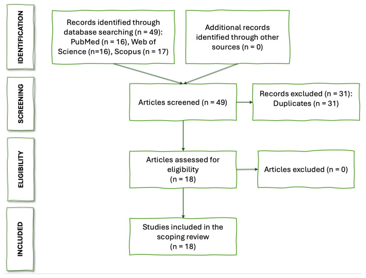
Micro-computed tomography (micro-CT) is a non-destructive imaging technique that offers highly detailed, 3D visualizations of a target specimen. In the context of breast cancer, micro-CT has emerged as a promising tool for analyzing microcalcifications (MCs), tiny calcium deposits that can indicate at an early stage the presence of cancer. This review aimed to explore the current applications of micro-CT in analyzing breast MCs (ex vivo, animal models, and phantoms) and to identify potential avenues in scientific research. We followed PRISMA guidelines for scoping reviews, yielding 18 studies that met our criteria. The studies varied in their purposes: feasibility and optimization of micro-CT for breast cancer imaging and MC analysis/diagnosis, comparison with other imaging modalities, development of micro-CT scanners and processing algorithms, enhancement of MC detection through contrast agents, etc. In conclusion, micro-CT offers superior image quality and detailed visualization of breast tissue (especially tumor masses and MCs), surpassing traditional methods like mammography and approaching the level of detail of histology. It holds great potential to enhance our understanding of MC characteristics and breast pathologies when used as a supplementary tool. Further research will solidify its role in clinical practice and potentially expand its applications in breast cancer studies.
TomographyMedicine-Radiology, Nuclear Medicine and Imaging
CiteScore
2.70
自引率
10.50%
发文量
222
期刊介绍:
TomographyTM publishes basic (technical and pre-clinical) and clinical scientific articles which involve the advancement of imaging technologies. Tomography encompasses studies that use single or multiple imaging modalities including for example CT, US, PET, SPECT, MR and hyperpolarization technologies, as well as optical modalities (i.e. bioluminescence, photoacoustic, endomicroscopy, fiber optic imaging and optical computed tomography) in basic sciences, engineering, preclinical and clinical medicine.
Tomography also welcomes studies involving exploration and refinement of contrast mechanisms and image-derived metrics within and across modalities toward the development of novel imaging probes for image-based feedback and intervention. The use of imaging in biology and medicine provides unparalleled opportunities to noninvasively interrogate tissues to obtain real-time dynamic and quantitative information required for diagnosis and response to interventions and to follow evolving pathological conditions. As multi-modal studies and the complexities of imaging technologies themselves are ever increasing to provide advanced information to scientists and clinicians.
Tomography provides a unique publication venue allowing investigators the opportunity to more precisely communicate integrated findings related to the diverse and heterogeneous features associated with underlying anatomical, physiological, functional, metabolic and molecular genetic activities of normal and diseased tissue. Thus Tomography publishes peer-reviewed articles which involve the broad use of imaging of any tissue and disease type including both preclinical and clinical investigations. In addition, hardware/software along with chemical and molecular probe advances are welcome as they are deemed to significantly contribute towards the long-term goal of improving the overall impact of imaging on scientific and clinical discovery.


 分享
分享
 求助内容:
求助内容: 应助结果提醒方式:
 扫码关注我们
