Brachial plexopathy due to perineural tumor spread: a retrospective single-center experience of clinical manifestations, diagnosis, treatments, and outcomes
{"title":"Brachial plexopathy due to perineural tumor spread: a retrospective single-center experience of clinical manifestations, diagnosis, treatments, and outcomes","authors":"Yu Jin Im, Young Cheol Yoon, Duk Hyun Sung","doi":"10.1007/s00701-024-06381-8","DOIUrl":null,"url":null,"abstract":"<div><h3>Background</h3><p>Perineural tumor spread (PNTS) to the brachial plexus (BP) is a rare and challenging condition. This study aimed to elucidate the clinical presentations, diagnostic challenges, and outcomes of patients with PNTS to the BP.</p><h3>Methods</h3><p>We retrospectively reviewed patients diagnosed with PNTS to the BP at our institution between January 2009 and June 2024. Clinical characteristics, magnetic resonance imaging (MRI), <sup>18</sup>F-fluorodeoxyglucose (<sup>18</sup>F-FDG) positron emission tomography/computed tomography (PET/CT) findings, and treatment outcomes were analyzed.</p><h3>Results</h3><p>Seven patients (mean age, 50.3 years) were identified. The primary cancer diagnoses included invasive ductal carcinoma of the breast (<i>n</i> = 3), metaplastic carcinoma of the breast (<i>n</i> = 1), lung adenocarcinoma (<i>n</i> = 2), and papillary thyroid carcinoma (<i>n</i> = 1). The median time from the initial cancer diagnosis to PNTS symptom onset was 71.0 months. All patients initially presented with progressive unilateral pain or paresthesia, followed by motor weakness. Lower trunk plexopathy was the most common electrodiagnostic finding (<i>n</i> = 5). In most patients, BP MRI showed diffuse tubular enlargement and T2 hyperintensity throughout the BP (<i>n</i> = 6), with gadolinium enhancement primarily in the proximal regions (<i>n</i> = 7). <sup>18</sup>F-FDG PET/CT demonstrated increased uptake in the BP, most prominently at the cervical spinal root or trunk levels (<i>n</i> = 6). Despite treatment, neurological outcomes were generally poor. Six of the seven patients died after a median follow-up of 19 months post-PNTS diagnosis.</p><h3>Conclusions</h3><p>PNTS to the BP can occur years after initial cancer diagnosis and may signify cancer progression. A high index of suspicion is crucial for timely diagnosis, particularly in patients with cancer and progressive upper extremity symptoms. Comprehensive imaging, including BP MRI and PET/CT, is essential for diagnosis. Despite treatment, prognosis remains poor, highlighting the need for improved diagnostic and therapeutic strategies.</p></div>","PeriodicalId":7370,"journal":{"name":"Acta Neurochirurgica","volume":"166 1","pages":""},"PeriodicalIF":1.9000,"publicationDate":"2024-12-02","publicationTypes":"Journal Article","fieldsOfStudy":null,"isOpenAccess":false,"openAccessPdf":"","citationCount":"0","resultStr":null,"platform":"Semanticscholar","paperid":null,"PeriodicalName":"Acta Neurochirurgica","FirstCategoryId":"3","ListUrlMain":"https://link.springer.com/article/10.1007/s00701-024-06381-8","RegionNum":3,"RegionCategory":"医学","ArticlePicture":[],"TitleCN":null,"AbstractTextCN":null,"PMCID":null,"EPubDate":"","PubModel":"","JCR":"Q3","JCRName":"CLINICAL NEUROLOGY","Score":null,"Total":0}
引用次数: 0
Abstract
Background
Perineural tumor spread (PNTS) to the brachial plexus (BP) is a rare and challenging condition. This study aimed to elucidate the clinical presentations, diagnostic challenges, and outcomes of patients with PNTS to the BP.
Methods
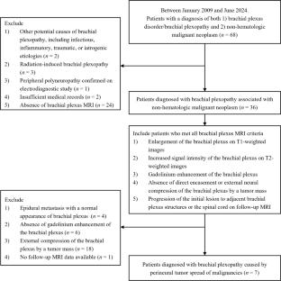
We retrospectively reviewed patients diagnosed with PNTS to the BP at our institution between January 2009 and June 2024. Clinical characteristics, magnetic resonance imaging (MRI), 18F-fluorodeoxyglucose (18F-FDG) positron emission tomography/computed tomography (PET/CT) findings, and treatment outcomes were analyzed.
Results
Seven patients (mean age, 50.3 years) were identified. The primary cancer diagnoses included invasive ductal carcinoma of the breast (n = 3), metaplastic carcinoma of the breast (n = 1), lung adenocarcinoma (n = 2), and papillary thyroid carcinoma (n = 1). The median time from the initial cancer diagnosis to PNTS symptom onset was 71.0 months. All patients initially presented with progressive unilateral pain or paresthesia, followed by motor weakness. Lower trunk plexopathy was the most common electrodiagnostic finding (n = 5). In most patients, BP MRI showed diffuse tubular enlargement and T2 hyperintensity throughout the BP (n = 6), with gadolinium enhancement primarily in the proximal regions (n = 7). 18F-FDG PET/CT demonstrated increased uptake in the BP, most prominently at the cervical spinal root or trunk levels (n = 6). Despite treatment, neurological outcomes were generally poor. Six of the seven patients died after a median follow-up of 19 months post-PNTS diagnosis.
Conclusions
PNTS to the BP can occur years after initial cancer diagnosis and may signify cancer progression. A high index of suspicion is crucial for timely diagnosis, particularly in patients with cancer and progressive upper extremity symptoms. Comprehensive imaging, including BP MRI and PET/CT, is essential for diagnosis. Despite treatment, prognosis remains poor, highlighting the need for improved diagnostic and therapeutic strategies.
期刊介绍:
The journal "Acta Neurochirurgica" publishes only original papers useful both to research and clinical work. Papers should deal with clinical neurosurgery - diagnosis and diagnostic techniques, operative surgery and results, postoperative treatment - or with research work in neuroscience if the underlying questions or the results are of neurosurgical interest. Reports on congresses are given in brief accounts. As official organ of the European Association of Neurosurgical Societies the journal publishes all announcements of the E.A.N.S. and reports on the activities of its member societies. Only contributions written in English will be accepted.


 分享
分享
 求助内容:
求助内容: 应助结果提醒方式:
 扫码关注我们
