Exposure to a 0.9-GHz electromagnetic field on postnatal days 21–45 may trigger the renin-angiotensin system in male rat: a histological and biochemical study
Ayşe İkinci Keleş, Haydar Kaya, Gökhan Keleş, Hüseyin Serkan Erol, Tolga Mercantepe, Ersan Odaci
{"title":"Exposure to a 0.9-GHz electromagnetic field on postnatal days 21–45 may trigger the renin-angiotensin system in male rat: a histological and biochemical study","authors":"Ayşe İkinci Keleş, Haydar Kaya, Gökhan Keleş, Hüseyin Serkan Erol, Tolga Mercantepe, Ersan Odaci","doi":"10.1007/s10735-024-10317-y","DOIUrl":null,"url":null,"abstract":"<div><p>The aim of this study was to examine the relationship between the renin-angiotensin system (RAS) and histological and biochemical changes occurring in the kidney tissue of male rats exposed to a 0.9 GHz electromagnetic field (EMF). Twelve male rats aged 21 days were randomly assigned to control (C-Gr) and EMF (EMF-Gr) groups. No procedure was performed on C-Gr, while the EMF-Gr rats were exposed to a 0.9 GHz EMF on postnatal days 21–45 (one hour a day for 25 days). Tissues were removed at the end of the experiment and evaluated using biochemical, and histopathological methods. Increased kidney tissue volume and weight and total body weight were determined in the group exposed to EMF. Lipid peroxidation, glutathione, catalase, and superoxide dismutase also increased in the kidney tissue of the EMF-Gr rats. Histopathological evaluation revealed cortical/medullary bleeding/obstruction and widespread fibrosis, dilatation, vacuolization, and degeneration in distal and proximal tubules, decreased and atypical parietal cells, and degeneration in epithelial cells. Additionally, dilated and degenerated glomeruli in the Malpighian body, Bowman’s membrane degeneration and degeneration in the vascular pole, podocyte, pedicel and mesangial cells were also observed. As a result of exposure to EMF, oxidative stress, tissue volume and weight increased, and histopathological changes caused the formation of a pathway that triggers RAS in kidney tissues. In conclusion, long-term exposure to 0.9 GHz EMF can activate the renin-angiotensin system in the rat kidney, and we think that such activation may be associated with structural, histopathological, and biochemical changes occurring in renal tissue.</p></div>","PeriodicalId":650,"journal":{"name":"Journal of Molecular Histology","volume":"56 1","pages":""},"PeriodicalIF":2.2000,"publicationDate":"2024-12-04","publicationTypes":"Journal Article","fieldsOfStudy":null,"isOpenAccess":false,"openAccessPdf":"","citationCount":"0","resultStr":null,"platform":"Semanticscholar","paperid":null,"PeriodicalName":"Journal of Molecular Histology","FirstCategoryId":"99","ListUrlMain":"https://link.springer.com/article/10.1007/s10735-024-10317-y","RegionNum":4,"RegionCategory":"生物学","ArticlePicture":[],"TitleCN":null,"AbstractTextCN":null,"PMCID":null,"EPubDate":"","PubModel":"","JCR":"Q3","JCRName":"CELL BIOLOGY","Score":null,"Total":0}
引用次数: 0
Abstract
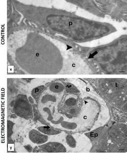
The aim of this study was to examine the relationship between the renin-angiotensin system (RAS) and histological and biochemical changes occurring in the kidney tissue of male rats exposed to a 0.9 GHz electromagnetic field (EMF). Twelve male rats aged 21 days were randomly assigned to control (C-Gr) and EMF (EMF-Gr) groups. No procedure was performed on C-Gr, while the EMF-Gr rats were exposed to a 0.9 GHz EMF on postnatal days 21–45 (one hour a day for 25 days). Tissues were removed at the end of the experiment and evaluated using biochemical, and histopathological methods. Increased kidney tissue volume and weight and total body weight were determined in the group exposed to EMF. Lipid peroxidation, glutathione, catalase, and superoxide dismutase also increased in the kidney tissue of the EMF-Gr rats. Histopathological evaluation revealed cortical/medullary bleeding/obstruction and widespread fibrosis, dilatation, vacuolization, and degeneration in distal and proximal tubules, decreased and atypical parietal cells, and degeneration in epithelial cells. Additionally, dilated and degenerated glomeruli in the Malpighian body, Bowman’s membrane degeneration and degeneration in the vascular pole, podocyte, pedicel and mesangial cells were also observed. As a result of exposure to EMF, oxidative stress, tissue volume and weight increased, and histopathological changes caused the formation of a pathway that triggers RAS in kidney tissues. In conclusion, long-term exposure to 0.9 GHz EMF can activate the renin-angiotensin system in the rat kidney, and we think that such activation may be associated with structural, histopathological, and biochemical changes occurring in renal tissue.
期刊介绍:
The Journal of Molecular Histology publishes results of original research on the localization and expression of molecules in animal cells, tissues and organs. Coverage includes studies describing novel cellular or ultrastructural distributions of molecules which provide insight into biochemical or physiological function, development, histologic structure and disease processes.
Major research themes of particular interest include:
- Cell-Cell and Cell-Matrix Interactions;
- Connective Tissues;
- Development and Disease;
- Neuroscience.
Please note that the Journal of Molecular Histology does not consider manuscripts dealing with the application of immunological or other probes on non-standard laboratory animal models unless the results are clearly of significant and general biological importance.
The Journal of Molecular Histology publishes full-length original research papers, review articles, short communications and letters to the editors. All manuscripts are typically reviewed by two independent referees. The Journal of Molecular Histology is a continuation of The Histochemical Journal.


 分享
分享
 求助内容:
求助内容: 应助结果提醒方式:
 扫码关注我们
