Bahar Kartal, Ebru Alimoğulları, Gökhan Akkurt, Mustafa Alimogulları, Sevil Çaylı
{"title":"The histological investigation of the effects of electromagnetic radiation on rat ovaries","authors":"Bahar Kartal, Ebru Alimoğulları, Gökhan Akkurt, Mustafa Alimogulları, Sevil Çaylı","doi":"10.1007/s10735-024-10319-w","DOIUrl":null,"url":null,"abstract":"<div><p>People are now exposed to higher levels of electromagnetic radiation (EMR) due to the widespread use of mobile phones in recent years. The possible effects of this exposure on human health are related to EMR. It has been suggested that exposure to EMR has serious effects on reproduction. The study aimed to investigate the impact of exposure to EMR (4.5 GB; 2600 MHz) emitted by mobile phones on rat ovaries. 18 adult female Wistar albino rats were used in the study, and the animals were divided into three groups (<i>n</i> = 6): control, stand-by, and dialing. For 8 weeks, the experimental groups were subjected to 4.5 GB EMR at 2600 MHz while on standby and making 10-min calls every hour. The rats in the control group received no exposure. Hematoxylin–eosin (H&E) staining of ovarian tissues was performed for histomorphological examinations. Additionally, immunoexpression of autophagy-related protein Beclin-1, apoptosis marker Caspase-3, ovarian reserve marker FSH, and oxidative stress marker iNOS were investigated in the rat ovaries. Microscopic examinations showed follicular degeneration in the ovaries of the rats in the stand-by and dialing groups. The immunoexpression of Beclin-1, Caspase-3, FSH, and iNOS was detected in granulosa cells and the corpus luteum in ovarian tissues obtained from the two EMR-exposed groups. There was a significant increase in the immunoexpression of Beclin-1 and Caspase-3 in the dialing group compared to the other two groups. Additionally, the iNOS and FSH expressions were increased in both EMR exposure groups compared to the control. Our results suggest that EMR exposure harms the ovaries, and autophagy and apoptosis are involved in this process.</p></div>","PeriodicalId":650,"journal":{"name":"Journal of Molecular Histology","volume":"56 1","pages":""},"PeriodicalIF":2.2000,"publicationDate":"2024-12-04","publicationTypes":"Journal Article","fieldsOfStudy":null,"isOpenAccess":false,"openAccessPdf":"","citationCount":"0","resultStr":null,"platform":"Semanticscholar","paperid":null,"PeriodicalName":"Journal of Molecular Histology","FirstCategoryId":"99","ListUrlMain":"https://link.springer.com/article/10.1007/s10735-024-10319-w","RegionNum":4,"RegionCategory":"生物学","ArticlePicture":[],"TitleCN":null,"AbstractTextCN":null,"PMCID":null,"EPubDate":"","PubModel":"","JCR":"Q3","JCRName":"CELL BIOLOGY","Score":null,"Total":0}
引用次数: 0
Abstract
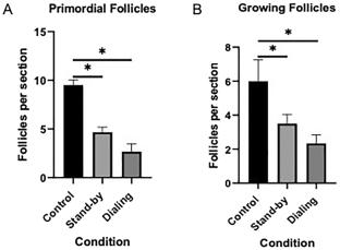
People are now exposed to higher levels of electromagnetic radiation (EMR) due to the widespread use of mobile phones in recent years. The possible effects of this exposure on human health are related to EMR. It has been suggested that exposure to EMR has serious effects on reproduction. The study aimed to investigate the impact of exposure to EMR (4.5 GB; 2600 MHz) emitted by mobile phones on rat ovaries. 18 adult female Wistar albino rats were used in the study, and the animals were divided into three groups (n = 6): control, stand-by, and dialing. For 8 weeks, the experimental groups were subjected to 4.5 GB EMR at 2600 MHz while on standby and making 10-min calls every hour. The rats in the control group received no exposure. Hematoxylin–eosin (H&E) staining of ovarian tissues was performed for histomorphological examinations. Additionally, immunoexpression of autophagy-related protein Beclin-1, apoptosis marker Caspase-3, ovarian reserve marker FSH, and oxidative stress marker iNOS were investigated in the rat ovaries. Microscopic examinations showed follicular degeneration in the ovaries of the rats in the stand-by and dialing groups. The immunoexpression of Beclin-1, Caspase-3, FSH, and iNOS was detected in granulosa cells and the corpus luteum in ovarian tissues obtained from the two EMR-exposed groups. There was a significant increase in the immunoexpression of Beclin-1 and Caspase-3 in the dialing group compared to the other two groups. Additionally, the iNOS and FSH expressions were increased in both EMR exposure groups compared to the control. Our results suggest that EMR exposure harms the ovaries, and autophagy and apoptosis are involved in this process.
期刊介绍:
The Journal of Molecular Histology publishes results of original research on the localization and expression of molecules in animal cells, tissues and organs. Coverage includes studies describing novel cellular or ultrastructural distributions of molecules which provide insight into biochemical or physiological function, development, histologic structure and disease processes.
Major research themes of particular interest include:
- Cell-Cell and Cell-Matrix Interactions;
- Connective Tissues;
- Development and Disease;
- Neuroscience.
Please note that the Journal of Molecular Histology does not consider manuscripts dealing with the application of immunological or other probes on non-standard laboratory animal models unless the results are clearly of significant and general biological importance.
The Journal of Molecular Histology publishes full-length original research papers, review articles, short communications and letters to the editors. All manuscripts are typically reviewed by two independent referees. The Journal of Molecular Histology is a continuation of The Histochemical Journal.


 分享
分享
 求助内容:
求助内容: 应助结果提醒方式:
 扫码关注我们
