Oscar Ariel Bautista, Vanessa Castro, Carolina Rodriguez, Eduardo Mastrangelo Marinho Falcão, Regina Casz Schechtman, Luna Azulay-Abulafia
{"title":"Trichoscopy of tinea capitis caused by Microsporum audouinii","authors":"Oscar Ariel Bautista, Vanessa Castro, Carolina Rodriguez, Eduardo Mastrangelo Marinho Falcão, Regina Casz Schechtman, Luna Azulay-Abulafia","doi":"10.1002/jvc2.530","DOIUrl":null,"url":null,"abstract":"<p><i>Tinea capitis</i> (TC), primarily affecting children aged 3–7, has globally increased in prevalence, and <i>Microsporum canis, a</i> zoophilic fungi, and <i>Trichophyton tonsurans, an</i> anthropophilic fungi, have become the major aetiologic agents.<span><sup>1</sup></span> In the 21st century, reports indicate an increase in the number of cases of anthropophilic transmission due to migrations, urbanization, changes in lifestyle and socioeconomic levels.<span><sup>1</sup></span></p><p><i>Microsporum audouinii</i>, an anthropophilic fungi endemic to Africa, causes milder inflammatory and chronic TC with late detection due to the lack of subjective symptoms, in comparison with <i>M. canis</i> infections which are more severe and accompanied by erythema.<span><sup>2</sup></span></p><p>In 2017, the first two cases of TC caused by <i>M. audouinii</i> in South America were reported in Brazil, involving two siblings without a history of prior travel, indicating possible autochthonous transmission.<span><sup>3</sup></span></p><p>Mycological examination is considered the gold standard diagnostic method of TC.<span><sup>4</sup></span> However, trichoscopy can be useful for making a correct and early diagnosis before culture results are available.<span><sup>4</sup></span> Trichoscopy exhibits higher sensitivity (94% vs. 49.1%) and specificity (83%) compared to direct KOH testing in diagnosing TC.<span><sup>5</sup></span></p><p>An observational, descriptive cross-sectional study was conducted at Instituto de Dermatologia Professor Rubem David Azulay of the Santa Casa da Misericórdia do Rio de Janeiro. We describe the trichoscopic findings of nine children, eight males and one female, aged between 3 and 14 years, average age 7 years, diagnosed with TC caused by <i>M. audouinii</i> from 2019 to 2023. Fluorescence with Wood lamp was performed identifying bluish-green fluorescence in all of them (Figure 1). Data were extracted from the patients' medical records to analyze clinical and sociodemographic characteristics. Statistical analysis involved calculating absolute frequencies (<i>n</i>) and relative frequencies (percentages).</p><p>The diagnosis of TC caused by <i>M. audouinii</i> was confirmed by direct KOH testing and culture. KOH testing showed ectothrix parasitism in all samples, while culture on Sabouraud agar displayed colonies of <i>Microsporum</i> spp. Slide cultures on potato dextrose agar were assessed under light microscopy using lactophenol cotton blue identifying species-specific structures. Growth pattern on boiled rice confirmed the identification. Trichoscopy performed without immersion liquid showed specific TC signs in at least two hairs per analyzed field, assessed by two independent dermatologists. Patients with recent antifungal medication use or inflammatory TC were excluded.</p><p>The characteristic trichoscopic findings evaluated for TC observed in the patients were as follows (<i>n</i> = 9): Morse code-like hairs in seven (78%), zigzag hairs in three (33%), whitish sheath in three (33%), comma hairs in three (33%), corkscrew hairs in one (11%) (Figure 2).</p><p>The common less specific trichoscopic findings of TC assessed were as follows (<i>n</i> = 9): diffuse scaling in nine (100%), perifollicular scaling in nine (100%), broken hairs in nine (100%) and black dots in 7/9 (78%) (Figure 2).</p><p>Slowinska et al. identified the comma hair as a distinctive marker for TC,<span><sup>6</sup></span> with subsequent studies describing other specific signs such as corkscrew, zigzag and Morse code-like hairs, as well as a whitish sheath.<span><sup>7, 8</sup></span> While broken hairs, black dots and scaling are commonly observed in TC, they are not disease-specific and may be seen in other scalp conditions.<span><sup>9</sup></span></p><p>Trichoscopy, detecting single specific signs, can predict TC and aid in early diagnosis and treatment initiation before culture results are available, thus reducing contagion risk.<span><sup>9</sup></span> Morse code-like hairs are highly specific for TC caused by <i>Microsporum</i>, suggesting species differentiation.<span><sup>10</sup></span></p><p>Trichoscopy combined with Wood's light fluorescence and epidemiological history, helps to suspect the type of parasitism and, consequently, the aetiological agent. This work is pioneering in the identification of <i>M. audouinii</i>, and the observation of specific trichoscopic findings, such as Morse code-like hairs identified in more than 50% of the samples, and common less especific findings like perifollicular and diffuse scaling, broken hairs and black dots, will help in suspecting this agent in TC patients, especially in areas where it has been shown to be emerging, such as Rio de Janeiro, enabling early treatment initiation before culture results and potentially reducing disease incidence.</p><p>All authors contributed to the study conception and design. Material preparation, data collection and analysis were performed by Luna Azulay-Abulafia, Regina Casz Schechtman, Eduardo Mastrangelo Marinho Falcão, Oscar Ariel Bautista, Vanessa Castro and Carolina Rodriguez. The first draft of the manuscript was written by Oscar Ariel Bautista and all authors commented on previous versions of the manuscript. All authors read and approved the final manuscript.</p><p>The authors declare no conflict of interest.</p><p>The parents/guardians of minor patients have given written informed consent for their child's participation in the study, as well as for the use of their child's deidentified, anonymized, aggregated data and case details (including photographs) for publication. This is an observational study. Approval was granted by the Ethics Committee of Pontifical Catholic University of Rio de Janeiro (PUC-Rio)/Rio de Janeiro, Brazil, 16 October 2023 (No. 81/2023).</p>","PeriodicalId":94325,"journal":{"name":"JEADV clinical practice","volume":"3 5","pages":"1698-1700"},"PeriodicalIF":0.5000,"publicationDate":"2024-08-21","publicationTypes":"Journal Article","fieldsOfStudy":null,"isOpenAccess":false,"openAccessPdf":"https://onlinelibrary.wiley.com/doi/epdf/10.1002/jvc2.530","citationCount":"0","resultStr":null,"platform":"Semanticscholar","paperid":null,"PeriodicalName":"JEADV clinical practice","FirstCategoryId":"1085","ListUrlMain":"https://onlinelibrary.wiley.com/doi/10.1002/jvc2.530","RegionNum":0,"RegionCategory":null,"ArticlePicture":[],"TitleCN":null,"AbstractTextCN":null,"PMCID":null,"EPubDate":"","PubModel":"","JCR":"","JCRName":"","Score":null,"Total":0}
引用次数: 0
Abstract
Tinea capitis (TC), primarily affecting children aged 3–7, has globally increased in prevalence, and Microsporum canis, a zoophilic fungi, and Trichophyton tonsurans, an anthropophilic fungi, have become the major aetiologic agents.1 In the 21st century, reports indicate an increase in the number of cases of anthropophilic transmission due to migrations, urbanization, changes in lifestyle and socioeconomic levels.1
Microsporum audouinii, an anthropophilic fungi endemic to Africa, causes milder inflammatory and chronic TC with late detection due to the lack of subjective symptoms, in comparison with M. canis infections which are more severe and accompanied by erythema.2
In 2017, the first two cases of TC caused by M. audouinii in South America were reported in Brazil, involving two siblings without a history of prior travel, indicating possible autochthonous transmission.3
Mycological examination is considered the gold standard diagnostic method of TC.4 However, trichoscopy can be useful for making a correct and early diagnosis before culture results are available.4 Trichoscopy exhibits higher sensitivity (94% vs. 49.1%) and specificity (83%) compared to direct KOH testing in diagnosing TC.5
An observational, descriptive cross-sectional study was conducted at Instituto de Dermatologia Professor Rubem David Azulay of the Santa Casa da Misericórdia do Rio de Janeiro. We describe the trichoscopic findings of nine children, eight males and one female, aged between 3 and 14 years, average age 7 years, diagnosed with TC caused by M. audouinii from 2019 to 2023. Fluorescence with Wood lamp was performed identifying bluish-green fluorescence in all of them (Figure 1). Data were extracted from the patients' medical records to analyze clinical and sociodemographic characteristics. Statistical analysis involved calculating absolute frequencies (n) and relative frequencies (percentages).
The diagnosis of TC caused by M. audouinii was confirmed by direct KOH testing and culture. KOH testing showed ectothrix parasitism in all samples, while culture on Sabouraud agar displayed colonies of Microsporum spp. Slide cultures on potato dextrose agar were assessed under light microscopy using lactophenol cotton blue identifying species-specific structures. Growth pattern on boiled rice confirmed the identification. Trichoscopy performed without immersion liquid showed specific TC signs in at least two hairs per analyzed field, assessed by two independent dermatologists. Patients with recent antifungal medication use or inflammatory TC were excluded.
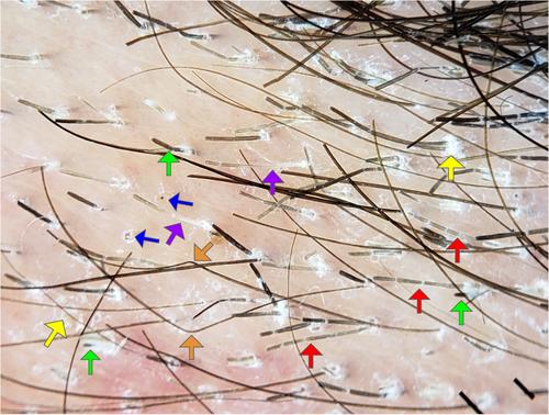
The characteristic trichoscopic findings evaluated for TC observed in the patients were as follows (n = 9): Morse code-like hairs in seven (78%), zigzag hairs in three (33%), whitish sheath in three (33%), comma hairs in three (33%), corkscrew hairs in one (11%) (Figure 2).
The common less specific trichoscopic findings of TC assessed were as follows (n = 9): diffuse scaling in nine (100%), perifollicular scaling in nine (100%), broken hairs in nine (100%) and black dots in 7/9 (78%) (Figure 2).
Slowinska et al. identified the comma hair as a distinctive marker for TC,6 with subsequent studies describing other specific signs such as corkscrew, zigzag and Morse code-like hairs, as well as a whitish sheath.7, 8 While broken hairs, black dots and scaling are commonly observed in TC, they are not disease-specific and may be seen in other scalp conditions.9
Trichoscopy, detecting single specific signs, can predict TC and aid in early diagnosis and treatment initiation before culture results are available, thus reducing contagion risk.9 Morse code-like hairs are highly specific for TC caused by Microsporum, suggesting species differentiation.10
Trichoscopy combined with Wood's light fluorescence and epidemiological history, helps to suspect the type of parasitism and, consequently, the aetiological agent. This work is pioneering in the identification of M. audouinii, and the observation of specific trichoscopic findings, such as Morse code-like hairs identified in more than 50% of the samples, and common less especific findings like perifollicular and diffuse scaling, broken hairs and black dots, will help in suspecting this agent in TC patients, especially in areas where it has been shown to be emerging, such as Rio de Janeiro, enabling early treatment initiation before culture results and potentially reducing disease incidence.
All authors contributed to the study conception and design. Material preparation, data collection and analysis were performed by Luna Azulay-Abulafia, Regina Casz Schechtman, Eduardo Mastrangelo Marinho Falcão, Oscar Ariel Bautista, Vanessa Castro and Carolina Rodriguez. The first draft of the manuscript was written by Oscar Ariel Bautista and all authors commented on previous versions of the manuscript. All authors read and approved the final manuscript.
The authors declare no conflict of interest.
The parents/guardians of minor patients have given written informed consent for their child's participation in the study, as well as for the use of their child's deidentified, anonymized, aggregated data and case details (including photographs) for publication. This is an observational study. Approval was granted by the Ethics Committee of Pontifical Catholic University of Rio de Janeiro (PUC-Rio)/Rio de Janeiro, Brazil, 16 October 2023 (No. 81/2023).


 分享
分享

 求助内容:
求助内容: 应助结果提醒方式:
 扫码关注我们
