Victoria A Shestakova, Ilya D Klabukov, Ilya V Kolobaev, Longfeng Rao, Dmitry A Atiakshin, Michael A Ignatyuk, Mikhail E Krasheninnikov, Bagavdin G Ahmedov, Sergey A Ivanov, Peter V Shegay, Andrey D Kaprin, Denis S Baranovskii
{"title":"Pathologically altered articular cartilage attracts intense chondrocyte invasion into the extracellular matrix: in vitro pilot study.","authors":"Victoria A Shestakova, Ilya D Klabukov, Ilya V Kolobaev, Longfeng Rao, Dmitry A Atiakshin, Michael A Ignatyuk, Mikhail E Krasheninnikov, Bagavdin G Ahmedov, Sergey A Ivanov, Peter V Shegay, Andrey D Kaprin, Denis S Baranovskii","doi":"10.1186/s43019-024-00249-y","DOIUrl":null,"url":null,"abstract":"<p><strong>Background: </strong>Due to non-vascularized and aneural structure, articular cartilage has limited self-repairing capacity. The aim of this study was to investigate the revitalization of inflammatory injured articular cartilage matrices by human nasal chondrocytes (hNC).</p><p><strong>Materials and methods: </strong>Cartilage matrix was prepared by devitalization of articular cartilage samples obtained intraoperatively from an adult patient undergoing knee joint replacement. hNC were obtained from native tissues by enzymatic digestion with further expansion over two passages. The obtained nasal chondrocytes were used to seed decellularized scaffolds, which were then cultured in vitro for 7, 14, or 21 days in chondrogenic medium. Migration was observed by histologic staining with fast green, safranin-O, and hematoxylin and scanning electron microscopy. Biochemical analysis was performed to determine the glycosaminoglycan (GAG) and DNA content of the cartilage using dimethylmethylene blue and CyQuant Cell Proliferation Assay Kit.</p><p><strong>Results: </strong>We seeded healthy and inflamed cartilage with nasal chondrocytes and found that the cells actively invade mainly pathologically altered cartilage. The results of biochemical quantitative analysis showed that the amount of DNA significantly increased by day 7 and decreased by day 14, while the quantitative values of GAGs had the opposite trend. Histological staining showed that cartilage formation occurred on day 7, intercellular spaces were filled with de novo synthesized cartilage matrix with significantly low GAG content on day 14, and newly formed GAG-rich cartilage was observed on day 21. The obtained data on cartilage regeneration were confirmed by scanning electron microscopy.</p><p><strong>Conclusions: </strong>Our preliminary results showed that human nasal chondrocytes are capable of infiltrating the pathologically altered extracellular matrix of articular cartilage damaged by arthritis, thereby promoting its repair to a physiologically relevant state.</p>","PeriodicalId":36317,"journal":{"name":"Knee Surgery and Related Research","volume":"36 1","pages":"42"},"PeriodicalIF":4.4000,"publicationDate":"2024-12-03","publicationTypes":"Journal Article","fieldsOfStudy":null,"isOpenAccess":false,"openAccessPdf":"https://www.ncbi.nlm.nih.gov/pmc/articles/PMC11613889/pdf/","citationCount":"0","resultStr":null,"platform":"Semanticscholar","paperid":null,"PeriodicalName":"Knee Surgery and Related Research","FirstCategoryId":"1085","ListUrlMain":"https://doi.org/10.1186/s43019-024-00249-y","RegionNum":0,"RegionCategory":null,"ArticlePicture":[],"TitleCN":null,"AbstractTextCN":null,"PMCID":null,"EPubDate":"","PubModel":"","JCR":"Q2","JCRName":"Medicine","Score":null,"Total":0}
引用次数: 0
Abstract
Background: Due to non-vascularized and aneural structure, articular cartilage has limited self-repairing capacity. The aim of this study was to investigate the revitalization of inflammatory injured articular cartilage matrices by human nasal chondrocytes (hNC).
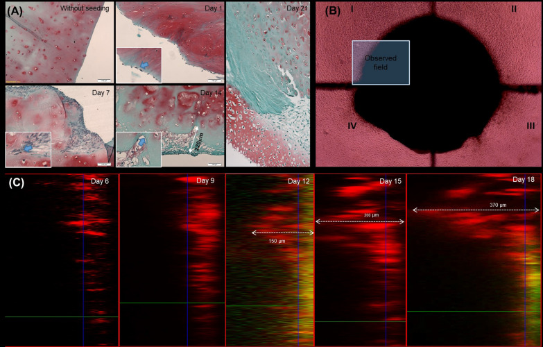
Materials and methods: Cartilage matrix was prepared by devitalization of articular cartilage samples obtained intraoperatively from an adult patient undergoing knee joint replacement. hNC were obtained from native tissues by enzymatic digestion with further expansion over two passages. The obtained nasal chondrocytes were used to seed decellularized scaffolds, which were then cultured in vitro for 7, 14, or 21 days in chondrogenic medium. Migration was observed by histologic staining with fast green, safranin-O, and hematoxylin and scanning electron microscopy. Biochemical analysis was performed to determine the glycosaminoglycan (GAG) and DNA content of the cartilage using dimethylmethylene blue and CyQuant Cell Proliferation Assay Kit.
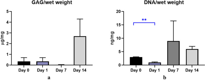
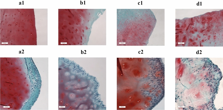
Results: We seeded healthy and inflamed cartilage with nasal chondrocytes and found that the cells actively invade mainly pathologically altered cartilage. The results of biochemical quantitative analysis showed that the amount of DNA significantly increased by day 7 and decreased by day 14, while the quantitative values of GAGs had the opposite trend. Histological staining showed that cartilage formation occurred on day 7, intercellular spaces were filled with de novo synthesized cartilage matrix with significantly low GAG content on day 14, and newly formed GAG-rich cartilage was observed on day 21. The obtained data on cartilage regeneration were confirmed by scanning electron microscopy.
Conclusions: Our preliminary results showed that human nasal chondrocytes are capable of infiltrating the pathologically altered extracellular matrix of articular cartilage damaged by arthritis, thereby promoting its repair to a physiologically relevant state.


 分享
分享
 求助内容:
求助内容: 应助结果提醒方式:
 扫码关注我们
