{"title":"Numerical, phenotypic and functional differences in human dermal resident memory T cells between reticular dermal layers.","authors":"Takuya Sato, Riko Asakawa, Youichi Ogawa, Yuka Nagasaka, Manao Kinoshita, Shinji Shimada, Akira Momosawa, Tatsuyoshi Kawamura","doi":"10.1002/ski2.457","DOIUrl":null,"url":null,"abstract":"<p><strong>Background: </strong>Resident memory T (T<sub>RM</sub>) cells and immunosuppressive Foxp3-expressing regulatory T<sub>RM</sub> (regT<sub>RM</sub>) cells are present in healthy, non-inflamed and inflamed human skin. Both types of cells are found in both the epidermis and dermis with the dermis being much thicker than the epidermis. However, it is unclear if T<sub>RM</sub> and regT<sub>RM</sub> cells differ between reticular dermal layers in terms of number, function or characteristics.</p><p><strong>Methods: </strong>This study examined numerical, phenotypic and functional differences in T<sub>RM</sub> and regT<sub>RM</sub> cells between the upper and lower reticular dermis in healthy, non-inflamed human skin using flow cytometry.</p><p><strong>Results: </strong>The phenotype and cytokine expressions of both types of cells were similar between reticular dermal layers. However, the cell count of both T<sub>RM</sub> and regT<sub>RM</sub> cells was significantly higher in the upper reticular dermis than in the lower reticular dermis.</p><p><strong>Conclusions: </strong>T<sub>RM</sub> and regT<sub>RM</sub> cells are phenotypically and functionally identical between reticular dermal layers. However, both types of cells are more abundant in the upper reticular dermis.</p>","PeriodicalId":74804,"journal":{"name":"Skin health and disease","volume":"4 6","pages":"e457"},"PeriodicalIF":0.0000,"publicationDate":"2024-12-01","publicationTypes":"Journal Article","fieldsOfStudy":null,"isOpenAccess":false,"openAccessPdf":"https://www.ncbi.nlm.nih.gov/pmc/articles/PMC11608865/pdf/","citationCount":"0","resultStr":null,"platform":"Semanticscholar","paperid":null,"PeriodicalName":"Skin health and disease","FirstCategoryId":"1085","ListUrlMain":"https://doi.org/10.1002/ski2.457","RegionNum":0,"RegionCategory":null,"ArticlePicture":[],"TitleCN":null,"AbstractTextCN":null,"PMCID":null,"EPubDate":"","PubModel":"","JCR":"Q3","JCRName":"Medicine","Score":null,"Total":0}
引用次数: 0
Abstract
Background: Resident memory T (TRM) cells and immunosuppressive Foxp3-expressing regulatory TRM (regTRM) cells are present in healthy, non-inflamed and inflamed human skin. Both types of cells are found in both the epidermis and dermis with the dermis being much thicker than the epidermis. However, it is unclear if TRM and regTRM cells differ between reticular dermal layers in terms of number, function or characteristics.
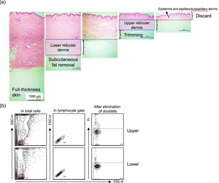
Methods: This study examined numerical, phenotypic and functional differences in TRM and regTRM cells between the upper and lower reticular dermis in healthy, non-inflamed human skin using flow cytometry.
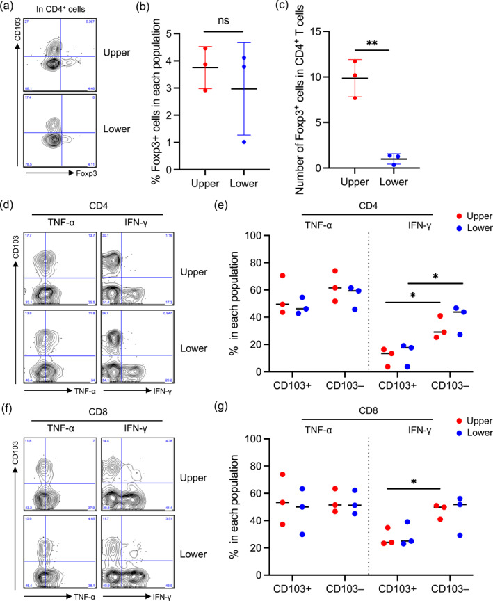
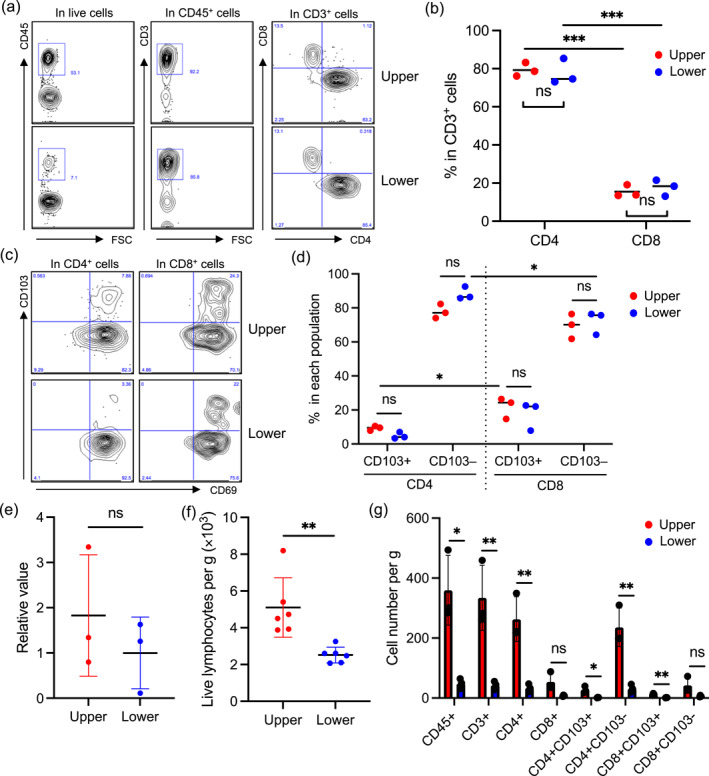
Results: The phenotype and cytokine expressions of both types of cells were similar between reticular dermal layers. However, the cell count of both TRM and regTRM cells was significantly higher in the upper reticular dermis than in the lower reticular dermis.
Conclusions: TRM and regTRM cells are phenotypically and functionally identical between reticular dermal layers. However, both types of cells are more abundant in the upper reticular dermis.


 分享
分享

 求助内容:
求助内容: 应助结果提醒方式:
 扫码关注我们
