Muhsin Ozgun Ozturk, Mustafa Resorlu, Sonay Aydin, Kemal Bugra Memis
{"title":"Use of the vertebrae and iliac bone as references for localizing the appendix vermiformis in computed tomography.","authors":"Muhsin Ozgun Ozturk, Mustafa Resorlu, Sonay Aydin, Kemal Bugra Memis","doi":"10.4329/wjr.v16.i11.629","DOIUrl":null,"url":null,"abstract":"<p><strong>Background: </strong>The appendix vermiformis is a part of the gastrointestinal tract, situated in the lower right quadrant of the abdomen. Acute appendicitis, acute inflammation of the appendix vermiformis, is the most common cause of acute abdomen requiring surgical intervention. Although computed tomography (CT) offers high diagnostic efficacy in assessing the appendix across various anatomical positions, it also involves radiation exposure. Reducing exposure factors and narrowing the field of view (FOV) are ways to decrease the radiation dose to the patient. To narrow the FOV, appendix locations within the population must be defined using metric markers.</p><p><strong>Aim: </strong>To determine the location of the appendix vermiformis on CT using the vertebrae and the right iliac bone as anatomical landmarks.</p><p><strong>Methods: </strong>This retrospective study examined 470 patients presenting with abdominal pain who underwent abdominal CT scans between January 01, 2015 and January 01, 2018. Forty-three patients were excluded due to various reasons. The most superior and inferior points and the origin of the appendix were measured separately in relation to the vertebrae and right iliac bone for localization. The population was divided into normal and acute appendicitis groups, and the relationship between appendix location and anthropometric parameters relationship was examined. <i>P</i> values below 0.05 were considered statistically significant.</p><p><strong>Results: </strong>The final analysis included 427 adult patients (206 females and 221 males) with a mean age of 42.1 ± 19.5 years. An ascending appendix course was the most common (90.4%). The appendix ranged from the L2 vertebral body level to the coccygeal vertebral level relative to the vertebrae. The appendix ranged between (-) 140.5 mm and (+) 87.4 mm relative to the right iliac bone. A negative correlation was found between patient age, height, body mass index, and the highest and lowest points of the appendix in regard to the vertebrae.</p><p><strong>Conclusion: </strong>The study's findings unveiled the locations of the appendix in the population in relation to the bony anatomical landmarks. These data can be used as the basis for future research aimed at reducing patient exposure to ionizing radiation.</p>","PeriodicalId":23819,"journal":{"name":"World journal of radiology","volume":"16 11","pages":"629-637"},"PeriodicalIF":1.2000,"publicationDate":"2024-11-28","publicationTypes":"Journal Article","fieldsOfStudy":null,"isOpenAccess":false,"openAccessPdf":"https://www.ncbi.nlm.nih.gov/pmc/articles/PMC11612806/pdf/","citationCount":"0","resultStr":null,"platform":"Semanticscholar","paperid":null,"PeriodicalName":"World journal of radiology","FirstCategoryId":"1085","ListUrlMain":"https://doi.org/10.4329/wjr.v16.i11.629","RegionNum":0,"RegionCategory":null,"ArticlePicture":[],"TitleCN":null,"AbstractTextCN":null,"PMCID":null,"EPubDate":"","PubModel":"","JCR":"Q3","JCRName":"RADIOLOGY, NUCLEAR MEDICINE & MEDICAL IMAGING","Score":null,"Total":0}
引用次数: 0
Abstract
Background: The appendix vermiformis is a part of the gastrointestinal tract, situated in the lower right quadrant of the abdomen. Acute appendicitis, acute inflammation of the appendix vermiformis, is the most common cause of acute abdomen requiring surgical intervention. Although computed tomography (CT) offers high diagnostic efficacy in assessing the appendix across various anatomical positions, it also involves radiation exposure. Reducing exposure factors and narrowing the field of view (FOV) are ways to decrease the radiation dose to the patient. To narrow the FOV, appendix locations within the population must be defined using metric markers.
Aim: To determine the location of the appendix vermiformis on CT using the vertebrae and the right iliac bone as anatomical landmarks.
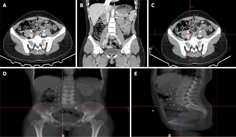
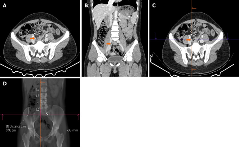
Methods: This retrospective study examined 470 patients presenting with abdominal pain who underwent abdominal CT scans between January 01, 2015 and January 01, 2018. Forty-three patients were excluded due to various reasons. The most superior and inferior points and the origin of the appendix were measured separately in relation to the vertebrae and right iliac bone for localization. The population was divided into normal and acute appendicitis groups, and the relationship between appendix location and anthropometric parameters relationship was examined. P values below 0.05 were considered statistically significant.
Results: The final analysis included 427 adult patients (206 females and 221 males) with a mean age of 42.1 ± 19.5 years. An ascending appendix course was the most common (90.4%). The appendix ranged from the L2 vertebral body level to the coccygeal vertebral level relative to the vertebrae. The appendix ranged between (-) 140.5 mm and (+) 87.4 mm relative to the right iliac bone. A negative correlation was found between patient age, height, body mass index, and the highest and lowest points of the appendix in regard to the vertebrae.
Conclusion: The study's findings unveiled the locations of the appendix in the population in relation to the bony anatomical landmarks. These data can be used as the basis for future research aimed at reducing patient exposure to ionizing radiation.


 分享
分享
 求助内容:
求助内容: 应助结果提醒方式:
 扫码关注我们
