Assessment of the physiological effects and safety of transpulmonary chemoembolization with doxorubicin on pulmonary tissue using a human-isolated lung perfusion model.
Alexis Slama, Hannah Steinberg, Stéphane Collaud, Özlem Okumus, Ralph-Axel Hilger, Sebastian Bauer, Hans-Ulrich Schildhaus, Clemens Aigner, Benedikt M Schaarschmidt
{"title":"Assessment of the physiological effects and safety of transpulmonary chemoembolization with doxorubicin on pulmonary tissue using a human-isolated lung perfusion model.","authors":"Alexis Slama, Hannah Steinberg, Stéphane Collaud, Özlem Okumus, Ralph-Axel Hilger, Sebastian Bauer, Hans-Ulrich Schildhaus, Clemens Aigner, Benedikt M Schaarschmidt","doi":"10.1186/s41747-024-00532-3","DOIUrl":null,"url":null,"abstract":"<p><strong>Background: </strong>Whole lung transpulmonary chemoembolization using a combination of doxorubicin (DXO) and degradable starch microspheres (DSM-TPCE) might be a promising treatment option in soft tissue sarcoma. To pave the way for clinical studies, this study aimed to evaluate the short-term effects of DSM-TPCE with DXO using an ex vivo isolated lung perfusion (ILP) model.</p><p><strong>Methods: </strong>Nine lung specimens retrieved from patients undergoing lobectomy underwent ex vivo ILP. In groups of three, lung specimens were either treated with sole DXO, sole DSM, or combined substances (DSM + DXO). During ex vivo ILP, histological samples were obtained from each lung every 15 min. Quantitative DXO analysis and histopathological grading of possible tissue damage using a five-point Likert scale was performed. Two-way repeated measures ANOVA tested for differences between treatment groups and changes over time.</p><p><strong>Results: </strong>We created a preclinical ex vivo ILP model to simulate the effects of DSM-TPCE. In histopathological analysis, only two specimens, treated with only DXO, showed an increase in parenchymal damage over time. No significant effect of time (3.3%, p = 0.305) or group (23.3; p = 0.331) was identified. Within the lung tissue, the DXO concentration ranged from 205 to 1,244 ng/g. No significant effects could be detected regarding different treatment groups (4.9% of total variation, p = 0.103).</p><p><strong>Conclusion: </strong>In an ex vivo ILP model using human lung lobes, the physiological effects of DSM-TPCE with DXO could be tested. Neither increased DXO concentrations in lung tissue nor histopathological changes indicating early lung toxicity were observed.</p><p><strong>Relevance statement: </strong>An ex vivo ILP model using human lung specimens did not show any signs of early lung toxicity after transpulmonary chemoembolization with DXO. These results support further evaluation of DSM-TPCE in phase I/II trials.</p><p><strong>Key points: </strong>Transpulmonary chemoembolization can be investigated in an ex vivo ILP model. DSM did not increase DXO in normal lung tissue. DSM did not increase parenchymal toxicity compared to the control groups.</p>","PeriodicalId":36926,"journal":{"name":"European Radiology Experimental","volume":"8 1","pages":"137"},"PeriodicalIF":3.6000,"publicationDate":"2024-12-05","publicationTypes":"Journal Article","fieldsOfStudy":null,"isOpenAccess":false,"openAccessPdf":"https://www.ncbi.nlm.nih.gov/pmc/articles/PMC11621295/pdf/","citationCount":"0","resultStr":null,"platform":"Semanticscholar","paperid":null,"PeriodicalName":"European Radiology Experimental","FirstCategoryId":"1085","ListUrlMain":"https://doi.org/10.1186/s41747-024-00532-3","RegionNum":0,"RegionCategory":null,"ArticlePicture":[],"TitleCN":null,"AbstractTextCN":null,"PMCID":null,"EPubDate":"","PubModel":"","JCR":"Q1","JCRName":"RADIOLOGY, NUCLEAR MEDICINE & MEDICAL IMAGING","Score":null,"Total":0}
引用次数: 0
Abstract
Background: Whole lung transpulmonary chemoembolization using a combination of doxorubicin (DXO) and degradable starch microspheres (DSM-TPCE) might be a promising treatment option in soft tissue sarcoma. To pave the way for clinical studies, this study aimed to evaluate the short-term effects of DSM-TPCE with DXO using an ex vivo isolated lung perfusion (ILP) model.
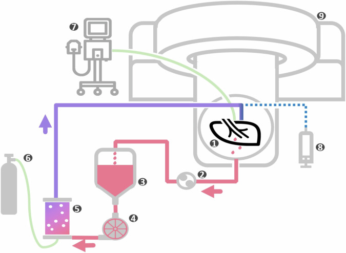
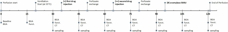
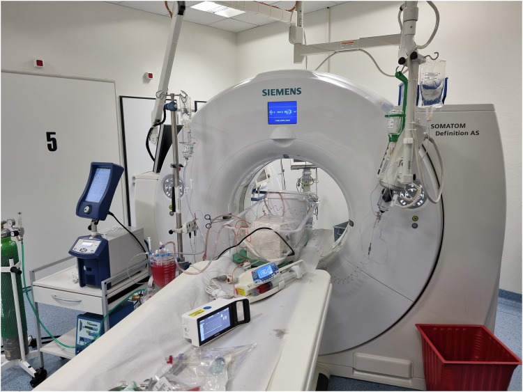
Methods: Nine lung specimens retrieved from patients undergoing lobectomy underwent ex vivo ILP. In groups of three, lung specimens were either treated with sole DXO, sole DSM, or combined substances (DSM + DXO). During ex vivo ILP, histological samples were obtained from each lung every 15 min. Quantitative DXO analysis and histopathological grading of possible tissue damage using a five-point Likert scale was performed. Two-way repeated measures ANOVA tested for differences between treatment groups and changes over time.
Results: We created a preclinical ex vivo ILP model to simulate the effects of DSM-TPCE. In histopathological analysis, only two specimens, treated with only DXO, showed an increase in parenchymal damage over time. No significant effect of time (3.3%, p = 0.305) or group (23.3; p = 0.331) was identified. Within the lung tissue, the DXO concentration ranged from 205 to 1,244 ng/g. No significant effects could be detected regarding different treatment groups (4.9% of total variation, p = 0.103).
Conclusion: In an ex vivo ILP model using human lung lobes, the physiological effects of DSM-TPCE with DXO could be tested. Neither increased DXO concentrations in lung tissue nor histopathological changes indicating early lung toxicity were observed.
Relevance statement: An ex vivo ILP model using human lung specimens did not show any signs of early lung toxicity after transpulmonary chemoembolization with DXO. These results support further evaluation of DSM-TPCE in phase I/II trials.
Key points: Transpulmonary chemoembolization can be investigated in an ex vivo ILP model. DSM did not increase DXO in normal lung tissue. DSM did not increase parenchymal toxicity compared to the control groups.


 分享
分享
 求助内容:
求助内容: 应助结果提醒方式:
 扫码关注我们
