Michał Bonczar, Jan Koszewski, Wiktor Czarnota, Martyna Dziedzic, Patryk Ostrowski, Kamil Możdżeń, Agnieszka Murawska, Paweł Hajdyła, Andrzej Walocha, Ewa Walocha, Jerzy Walocha, Mateusz Koziej
{"title":"The morphology of the lumbar vertebrae: a systematic review with meta-analysis of 1481 individuals with implications for spine surgery.","authors":"Michał Bonczar, Jan Koszewski, Wiktor Czarnota, Martyna Dziedzic, Patryk Ostrowski, Kamil Możdżeń, Agnieszka Murawska, Paweł Hajdyła, Andrzej Walocha, Ewa Walocha, Jerzy Walocha, Mateusz Koziej","doi":"10.1007/s00276-024-03509-4","DOIUrl":null,"url":null,"abstract":"<p><strong>Introduction: </strong>The aim of the present meta-analysis was to provide the most up-to-date and evidence-based results regarding the morphometric properties of the lumbar vertebrae.</p><p><strong>Methods: </strong>Medical databases such as PubMed, Scopus, Embase, Web of Science, Google Scholar, and Cochrane Library were searched through.</p><p><strong>Results: </strong>The results of this meta-analysis were established based on a total of 1481 patients. New results were established in 27 categories for each lumbar vertebra separately. The findings from this study reveal that the width of the spinal canal progressively increases towards the lower end of the lumbar spine (L1 = 22.04 mm, L5 = 26.46 mm). Additionally, the transverse processes exhibit a similar trend, widening as they approach the lower lumbar vertebrae (L1 = 68.08 mm, L5 = 85.91 mm). The pedicle height decreased from L1 to L4, with an increase observed at L5 (14.73 mm). No significant differences were observed between the measurements of the left and right pedicles.</p><p><strong>Conclusion: </strong>The presented results provide physicians with normative morphometric data on the lumbar vertebrae. Having adequate knowledge of the anatomy of the lumbar vertebrae may be of immense use for surgeons performing various spinal surgeries, such as pedicle screw fixation, percutaneous endoscopic transforaminal discectomy, or lumbar disc replacement.</p>","PeriodicalId":49461,"journal":{"name":"Surgical and Radiologic Anatomy","volume":"47 1","pages":"22"},"PeriodicalIF":1.2000,"publicationDate":"2024-12-06","publicationTypes":"Journal Article","fieldsOfStudy":null,"isOpenAccess":false,"openAccessPdf":"https://www.ncbi.nlm.nih.gov/pmc/articles/PMC11624215/pdf/","citationCount":"0","resultStr":null,"platform":"Semanticscholar","paperid":null,"PeriodicalName":"Surgical and Radiologic Anatomy","FirstCategoryId":"3","ListUrlMain":"https://doi.org/10.1007/s00276-024-03509-4","RegionNum":4,"RegionCategory":"医学","ArticlePicture":[],"TitleCN":null,"AbstractTextCN":null,"PMCID":null,"EPubDate":"","PubModel":"","JCR":"Q2","JCRName":"Medicine","Score":null,"Total":0}
引用次数: 0
Abstract
Introduction: The aim of the present meta-analysis was to provide the most up-to-date and evidence-based results regarding the morphometric properties of the lumbar vertebrae.
Methods: Medical databases such as PubMed, Scopus, Embase, Web of Science, Google Scholar, and Cochrane Library were searched through.
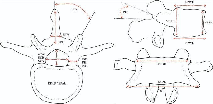
Results: The results of this meta-analysis were established based on a total of 1481 patients. New results were established in 27 categories for each lumbar vertebra separately. The findings from this study reveal that the width of the spinal canal progressively increases towards the lower end of the lumbar spine (L1 = 22.04 mm, L5 = 26.46 mm). Additionally, the transverse processes exhibit a similar trend, widening as they approach the lower lumbar vertebrae (L1 = 68.08 mm, L5 = 85.91 mm). The pedicle height decreased from L1 to L4, with an increase observed at L5 (14.73 mm). No significant differences were observed between the measurements of the left and right pedicles.
Conclusion: The presented results provide physicians with normative morphometric data on the lumbar vertebrae. Having adequate knowledge of the anatomy of the lumbar vertebrae may be of immense use for surgeons performing various spinal surgeries, such as pedicle screw fixation, percutaneous endoscopic transforaminal discectomy, or lumbar disc replacement.
期刊介绍:
Anatomy is a morphological science which cannot fail to interest the clinician. The practical application of anatomical research to clinical problems necessitates special adaptation and selectivity in choosing from numerous international works. Although there is a tendency to believe that meaningful advances in anatomy are unlikely, constant revision is necessary. Surgical and Radiologic Anatomy, the first international journal of Clinical anatomy has been created in this spirit.
Its goal is to serve clinicians, regardless of speciality-physicians, surgeons, radiologists or other specialists-as an indispensable aid with which they can improve their knowledge of anatomy. Each issue includes: Original papers, review articles, articles on the anatomical bases of medical, surgical and radiological techniques, articles of normal radiologic anatomy, brief reviews of anatomical publications of clinical interest.
Particular attention is given to high quality illustrations, which are indispensable for a better understanding of anatomical problems.
Surgical and Radiologic Anatomy is a journal written by anatomists for clinicians with a special interest in anatomy.


 分享
分享
 求助内容:
求助内容: 应助结果提醒方式:
 扫码关注我们
