Tianying Zheng, Yajing Zhu, Yidi Chen, Shengshi Mai, Lixin Xu, Hanyu Jiang, Ting Duan, Yuanan Wu, Yali Qu, Yinan Chen, Bin Song
{"title":"Fully automated MRI-based convolutional neural network for noninvasive diagnosis of cirrhosis.","authors":"Tianying Zheng, Yajing Zhu, Yidi Chen, Shengshi Mai, Lixin Xu, Hanyu Jiang, Ting Duan, Yuanan Wu, Yali Qu, Yinan Chen, Bin Song","doi":"10.1186/s13244-024-01872-9","DOIUrl":null,"url":null,"abstract":"<p><strong>Objectives: </strong>To develop and externally validate a fully automated diagnostic convolutional neural network (CNN) model for cirrhosis based on liver MRI and serum biomarkers.</p><p><strong>Methods: </strong>This multicenter retrospective study included consecutive patients receiving pathological evaluation of liver fibrosis stage and contrast-enhanced liver MRI between March 2010 and January 2024. On the training dataset, an MRI-based CNN model was constructed for cirrhosis against pathology, and then a combined model was developed integrating the CNN model and serum biomarkers. On the testing datasets, the area under the receiver operating characteristic curve (AUC) was computed to compare the diagnostic performance of the combined model with that of aminotransferase-to-platelet ratio index (APRI), fibrosis-4 index (FIB-4), and radiologists. The influence of potential confounders on the diagnostic performance was evaluated by subgroup analyses.</p><p><strong>Results: </strong>A total of 1315 patients (median age, 54 years; 1065 men; training, n = 840) were included, 855 (65%) with pathological cirrhosis. The CNN model was constructed on pre-contrast T1- and T2-weighted imaging, and the combined model was developed integrating the CNN model, age, and eight serum biomarkers. On the external testing dataset, the combined model achieved an AUC of 0.86, which outperformed FIB-4, APRI and two radiologists (AUC: 0.67 to 0.73, all p < 0.05). Subgroup analyses revealed comparable diagnostic performances of the combined model in patients with different sizes of focal liver lesions.</p><p><strong>Conclusion: </strong>Based on pre-contrast T1- and T2-weighted imaging, age, and serum biomarkers, the combined model allowed diagnosis of cirrhosis with moderate accuracy, independent of the size of focal liver lesions.</p><p><strong>Critical relevance statement: </strong>The fully automated convolutional neural network model utilizing pre-contrast MR imaging, age and serum biomarkers demonstrated moderate accuracy, outperforming FIB-4, APRI, and radiologists, independent of size of focal liver lesions, potentially facilitating noninvasive diagnosis of cirrhosis pending further validation.</p><p><strong>Key points: </strong>This fully automated convolutional neural network (CNN) model, using pre-contrast MRI, age, and serum biomarkers, diagnoses cirrhosis. The CNN model demonstrated an external testing dataset AUC of 0.86, independent of the size of focal liver lesions. The CNN model outperformed aminotransferase-to-platelet ratio index, fibrosis-4 index, and radiologists, potentially facilitating noninvasive diagnosis of cirrhosis.</p>","PeriodicalId":13639,"journal":{"name":"Insights into Imaging","volume":"15 1","pages":"298"},"PeriodicalIF":4.5000,"publicationDate":"2024-12-12","publicationTypes":"Journal Article","fieldsOfStudy":null,"isOpenAccess":false,"openAccessPdf":"https://www.ncbi.nlm.nih.gov/pmc/articles/PMC11638457/pdf/","citationCount":"0","resultStr":null,"platform":"Semanticscholar","paperid":null,"PeriodicalName":"Insights into Imaging","FirstCategoryId":"3","ListUrlMain":"https://doi.org/10.1186/s13244-024-01872-9","RegionNum":2,"RegionCategory":"医学","ArticlePicture":[],"TitleCN":null,"AbstractTextCN":null,"PMCID":null,"EPubDate":"","PubModel":"","JCR":"Q1","JCRName":"RADIOLOGY, NUCLEAR MEDICINE & MEDICAL IMAGING","Score":null,"Total":0}
引用次数: 0
Abstract
Objectives: To develop and externally validate a fully automated diagnostic convolutional neural network (CNN) model for cirrhosis based on liver MRI and serum biomarkers.
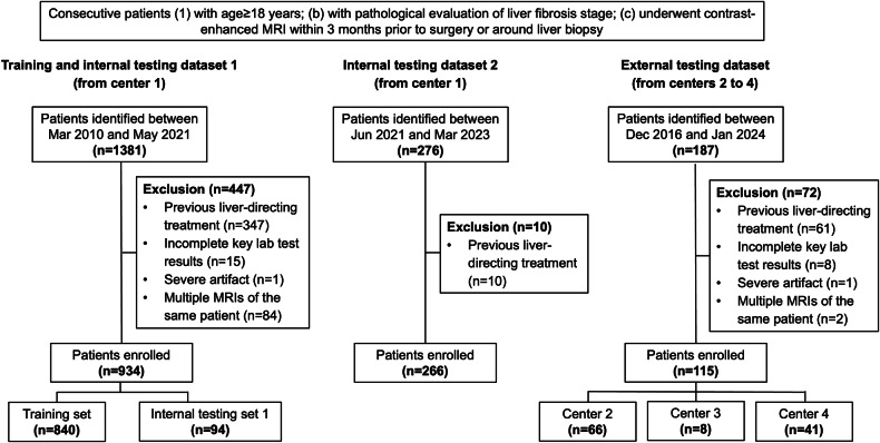
Methods: This multicenter retrospective study included consecutive patients receiving pathological evaluation of liver fibrosis stage and contrast-enhanced liver MRI between March 2010 and January 2024. On the training dataset, an MRI-based CNN model was constructed for cirrhosis against pathology, and then a combined model was developed integrating the CNN model and serum biomarkers. On the testing datasets, the area under the receiver operating characteristic curve (AUC) was computed to compare the diagnostic performance of the combined model with that of aminotransferase-to-platelet ratio index (APRI), fibrosis-4 index (FIB-4), and radiologists. The influence of potential confounders on the diagnostic performance was evaluated by subgroup analyses.
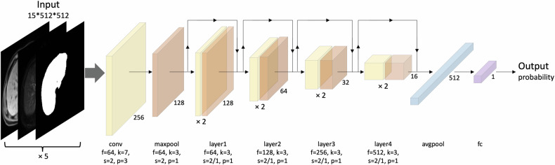
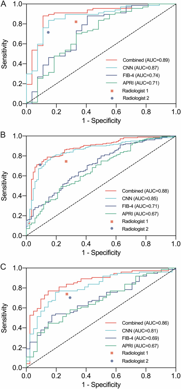
Results: A total of 1315 patients (median age, 54 years; 1065 men; training, n = 840) were included, 855 (65%) with pathological cirrhosis. The CNN model was constructed on pre-contrast T1- and T2-weighted imaging, and the combined model was developed integrating the CNN model, age, and eight serum biomarkers. On the external testing dataset, the combined model achieved an AUC of 0.86, which outperformed FIB-4, APRI and two radiologists (AUC: 0.67 to 0.73, all p < 0.05). Subgroup analyses revealed comparable diagnostic performances of the combined model in patients with different sizes of focal liver lesions.
Conclusion: Based on pre-contrast T1- and T2-weighted imaging, age, and serum biomarkers, the combined model allowed diagnosis of cirrhosis with moderate accuracy, independent of the size of focal liver lesions.
Critical relevance statement: The fully automated convolutional neural network model utilizing pre-contrast MR imaging, age and serum biomarkers demonstrated moderate accuracy, outperforming FIB-4, APRI, and radiologists, independent of size of focal liver lesions, potentially facilitating noninvasive diagnosis of cirrhosis pending further validation.
Key points: This fully automated convolutional neural network (CNN) model, using pre-contrast MRI, age, and serum biomarkers, diagnoses cirrhosis. The CNN model demonstrated an external testing dataset AUC of 0.86, independent of the size of focal liver lesions. The CNN model outperformed aminotransferase-to-platelet ratio index, fibrosis-4 index, and radiologists, potentially facilitating noninvasive diagnosis of cirrhosis.
期刊介绍:
Insights into Imaging (I³) is a peer-reviewed open access journal published under the brand SpringerOpen. All content published in the journal is freely available online to anyone, anywhere!
I³ continuously updates scientific knowledge and progress in best-practice standards in radiology through the publication of original articles and state-of-the-art reviews and opinions, along with recommendations and statements from the leading radiological societies in Europe.
Founded by the European Society of Radiology (ESR), I³ creates a platform for educational material, guidelines and recommendations, and a forum for topics of controversy.
A balanced combination of review articles, original papers, short communications from European radiological congresses and information on society matters makes I³ an indispensable source for current information in this field.
I³ is owned by the ESR, however authors retain copyright to their article according to the Creative Commons Attribution License (see Copyright and License Agreement). All articles can be read, redistributed and reused for free, as long as the author of the original work is cited properly.
The open access fees (article-processing charges) for this journal are kindly sponsored by ESR for all Members.
The journal went open access in 2012, which means that all articles published since then are freely available online.


 分享
分享
 求助内容:
求助内容: 应助结果提醒方式:
 扫码关注我们
