Marcony R Santhiago, Claudia R Morgado, Ellen Koo, Geetha Iyer, Bhaskar Srinivasan, Ruben Berrospi, Ramon Ghanem
{"title":"Corneal scar after ulcer in a young patient demanding visual restoration in a timely fashion treated with PTK + topo-guided PRK.","authors":"Marcony R Santhiago, Claudia R Morgado, Ellen Koo, Geetha Iyer, Bhaskar Srinivasan, Ruben Berrospi, Ramon Ghanem","doi":"10.1097/j.jcrs.0000000000001570","DOIUrl":null,"url":null,"abstract":"<p><p>A 23-year-old woman was referred for low visual acuity in the left eye after a corneal ulcer associated with contact lens use 2 years previously. The patient had a history of contact lens use, reported use of antibiotic eye drops with improvement of infection, and subsequent scarring. There were no comorbidities. The manifest refraction was -3.25 -2.25 × 180 (20/20) in the right eye and was -2.00 esf -2.00 × 165 (20/80) in the left eye. The patient demands a solution in a reasonable time because of the need for functional vision and possible restoration of her binocular functions. The slitlamp examination revealed a corneal scar partially affecting the visual axis (Figure 1JOURNAL/jcrs/04.03/02158034-202412000-00016/figure1/v/2024-12-12T192825Z/r/image-tiff). Corneal topography revealed an irregular pattern and spectral-domain optical coherence tomography (OCT) examinations revealed scarring in the anterior stroma (Figures 2 and 3JOURNAL/jcrs/04.03/02158034-202412000-00016/figure2/v/2024-12-12T192825Z/r/image-tiffJOURNAL/jcrs/04.03/02158034-202412000-00016/figure3/v/2024-12-12T192825Z/r/image-tiff). Given the patient's refraction, corneal scar, and visual demands, would you perform photorefractive keratectomy (PRK) treatment to correct ametropia and partially remove the anterior stroma? Would you perform excimer laser treatment for therapeutic purposes guided by topography? Would you opt for a 2-stage treatment, regularizing the cornea with neutral phototherapeutic keratectomy (PTK) or PRK treatment guided by topography and then correcting the ametropia? Considering the OTC maps, would you perform a femtosecond laser-assisted anterior lamellar keratoplasty (FALK), deep anterior lamellar keratoplasty (DALK), or even penetrating keratoplasty? Would you consider any other surgical step to prevent delayed cornea healing-persistent epithelial defect?</p>","PeriodicalId":15214,"journal":{"name":"Journal of cataract and refractive surgery","volume":"50 12","pages":"1293"},"PeriodicalIF":3.2000,"publicationDate":"2024-12-01","publicationTypes":"Journal Article","fieldsOfStudy":null,"isOpenAccess":false,"openAccessPdf":"https://www.ncbi.nlm.nih.gov/pmc/articles/PMC11556845/pdf/","citationCount":"0","resultStr":null,"platform":"Semanticscholar","paperid":null,"PeriodicalName":"Journal of cataract and refractive surgery","FirstCategoryId":"3","ListUrlMain":"https://doi.org/10.1097/j.jcrs.0000000000001570","RegionNum":3,"RegionCategory":"医学","ArticlePicture":[],"TitleCN":null,"AbstractTextCN":null,"PMCID":null,"EPubDate":"","PubModel":"","JCR":"Q2","JCRName":"OPHTHALMOLOGY","Score":null,"Total":0}
引用次数: 0
Abstract
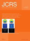
A 23-year-old woman was referred for low visual acuity in the left eye after a corneal ulcer associated with contact lens use 2 years previously. The patient had a history of contact lens use, reported use of antibiotic eye drops with improvement of infection, and subsequent scarring. There were no comorbidities. The manifest refraction was -3.25 -2.25 × 180 (20/20) in the right eye and was -2.00 esf -2.00 × 165 (20/80) in the left eye. The patient demands a solution in a reasonable time because of the need for functional vision and possible restoration of her binocular functions. The slitlamp examination revealed a corneal scar partially affecting the visual axis (Figure 1JOURNAL/jcrs/04.03/02158034-202412000-00016/figure1/v/2024-12-12T192825Z/r/image-tiff). Corneal topography revealed an irregular pattern and spectral-domain optical coherence tomography (OCT) examinations revealed scarring in the anterior stroma (Figures 2 and 3JOURNAL/jcrs/04.03/02158034-202412000-00016/figure2/v/2024-12-12T192825Z/r/image-tiffJOURNAL/jcrs/04.03/02158034-202412000-00016/figure3/v/2024-12-12T192825Z/r/image-tiff). Given the patient's refraction, corneal scar, and visual demands, would you perform photorefractive keratectomy (PRK) treatment to correct ametropia and partially remove the anterior stroma? Would you perform excimer laser treatment for therapeutic purposes guided by topography? Would you opt for a 2-stage treatment, regularizing the cornea with neutral phototherapeutic keratectomy (PTK) or PRK treatment guided by topography and then correcting the ametropia? Considering the OTC maps, would you perform a femtosecond laser-assisted anterior lamellar keratoplasty (FALK), deep anterior lamellar keratoplasty (DALK), or even penetrating keratoplasty? Would you consider any other surgical step to prevent delayed cornea healing-persistent epithelial defect?
期刊介绍:
The Journal of Cataract & Refractive Surgery (JCRS), a preeminent peer-reviewed monthly ophthalmology publication, is the official journal of the American Society of Cataract and Refractive Surgery (ASCRS) and the European Society of Cataract and Refractive Surgeons (ESCRS).
JCRS publishes high quality articles on all aspects of anterior segment surgery. In addition to original clinical studies, the journal features a consultation section, practical techniques, important cases, and reviews as well as basic science articles.


 分享
分享
 求助内容:
求助内容: 应助结果提醒方式:
 扫码关注我们
