Aida Glembocki, Robert Siddaway, Anthony Arnoldo, Molly Jakeman, Anthea Lafreniere
{"title":"Clinical and Pathological Features of a Schwannoma Harboring a <i>SH3PXD2A::HTRA1</i> Gene Fusion in a Pre-pubescent Patient.","authors":"Aida Glembocki, Robert Siddaway, Anthony Arnoldo, Molly Jakeman, Anthea Lafreniere","doi":"10.1177/10935266241308946","DOIUrl":null,"url":null,"abstract":"<p><p>An 11-year-old girl presented with a soft tissue lesion on the dorsal aspect of the left middle finger. Ultrasound imaging demonstrated a 2.8 cm × 0.8 cm × 0.8 cm lesion overlying the dorsal aspect of the base of the digit near the metacarpophalangeal joint. The patient's past medical history is remarkable for neuroblastoma, diagnosed at 9 months of age, with no MYCN amplification or 1p loss. We report a pediatric schwannoma harbouring a <i>SH3PXD2A::HTRA1</i> gene fusion with a distinctive serpentine histology. The lesion consisted of well-circumscribed nodules surrounded by thin EMA-positive perineural capsules. Each nodule was composed of lesional cells arranged in short fascicles with occasional clefting and a distinct \"serpentine\" palisading pattern. The lesion demonstrated Antoni A regions with Verocay body formation. No significant Antoni B areas were seen. The lesional Schwannian cells were bland with elongated and tapered nuclei, showing strong and diffuse positivity for S100. This pre-pubescent girl (Tanner Stage 2) is currently the youngest reported case of fusion-positive schwannoma. In addition, she has a significant prior history of a malignant neoplasm, and the lesion arose in an appendicular location.</p>","PeriodicalId":54634,"journal":{"name":"Pediatric and Developmental Pathology","volume":" ","pages":"137-141"},"PeriodicalIF":1.3000,"publicationDate":"2025-03-01","publicationTypes":"Journal Article","fieldsOfStudy":null,"isOpenAccess":false,"openAccessPdf":"https://www.ncbi.nlm.nih.gov/pmc/articles/PMC11894906/pdf/","citationCount":"0","resultStr":null,"platform":"Semanticscholar","paperid":null,"PeriodicalName":"Pediatric and Developmental Pathology","FirstCategoryId":"3","ListUrlMain":"https://doi.org/10.1177/10935266241308946","RegionNum":4,"RegionCategory":"医学","ArticlePicture":[],"TitleCN":null,"AbstractTextCN":null,"PMCID":null,"EPubDate":"2024/12/24 0:00:00","PubModel":"Epub","JCR":"Q3","JCRName":"PATHOLOGY","Score":null,"Total":0}
引用次数: 0
Abstract
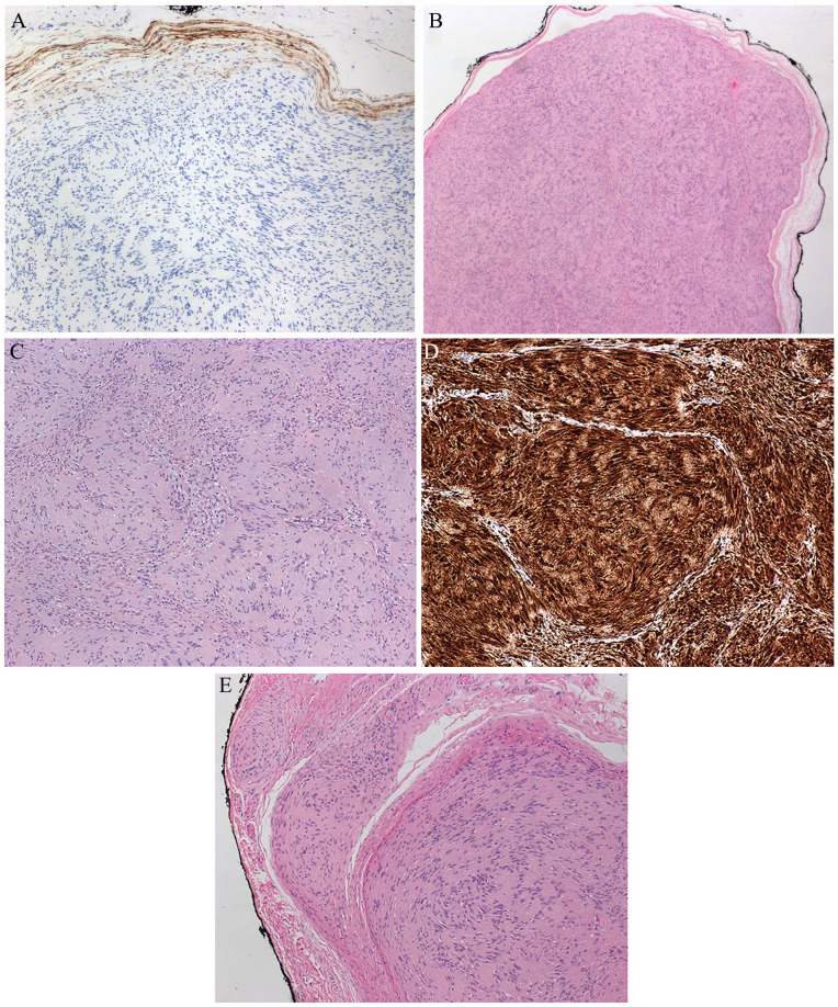
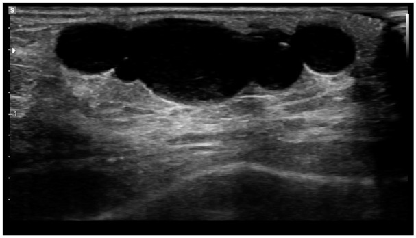
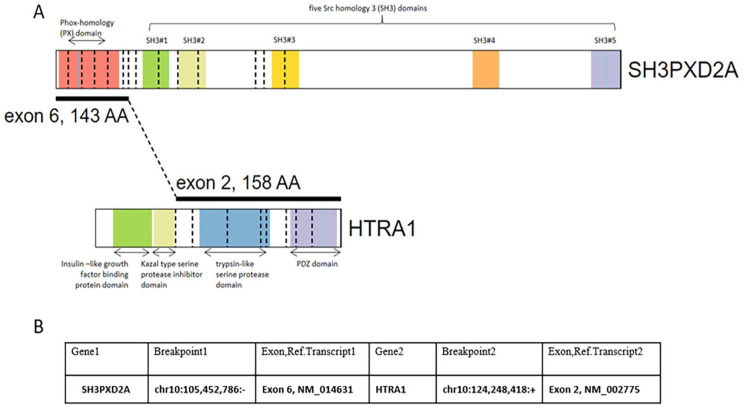
An 11-year-old girl presented with a soft tissue lesion on the dorsal aspect of the left middle finger. Ultrasound imaging demonstrated a 2.8 cm × 0.8 cm × 0.8 cm lesion overlying the dorsal aspect of the base of the digit near the metacarpophalangeal joint. The patient's past medical history is remarkable for neuroblastoma, diagnosed at 9 months of age, with no MYCN amplification or 1p loss. We report a pediatric schwannoma harbouring a SH3PXD2A::HTRA1 gene fusion with a distinctive serpentine histology. The lesion consisted of well-circumscribed nodules surrounded by thin EMA-positive perineural capsules. Each nodule was composed of lesional cells arranged in short fascicles with occasional clefting and a distinct "serpentine" palisading pattern. The lesion demonstrated Antoni A regions with Verocay body formation. No significant Antoni B areas were seen. The lesional Schwannian cells were bland with elongated and tapered nuclei, showing strong and diffuse positivity for S100. This pre-pubescent girl (Tanner Stage 2) is currently the youngest reported case of fusion-positive schwannoma. In addition, she has a significant prior history of a malignant neoplasm, and the lesion arose in an appendicular location.
一个11岁的女孩提出了软组织病变在背侧的左中指。超声成像显示一个2.8 cm × 0.8 cm × 0.8 cm的病变,位于手指基部背侧靠近掌指关节处。患者既往有神经母细胞瘤病史,9个月大时确诊,无MYCN扩增或1p缺失。我们报道了一例儿童神经鞘瘤,其中SH3PXD2A::HTRA1基因融合具有独特的蛇形组织学。病变包括边界清晰的结节,周围是薄的ema阳性的神经周围囊。每个结节由病变细胞组成,排列成短束状,偶有裂隙,呈明显的“蛇形”栅栏状。病变表现为Antoni A区,伴Verocay体形成。未见明显的Antoni B区。病变许旺氏细胞呈淡色,细胞核伸长、变细,S100呈强弥漫性阳性。这个青春期前的女孩(Tanner期2)是目前报道的最年轻的融合阳性神经鞘瘤病例。此外,她有明显的恶性肿瘤病史,病变发生在阑尾部位。
期刊介绍:
The Journal covers the spectrum of disorders of early development (including embryology, placentology, and teratology), gestational and perinatal diseases, and all diseases of childhood. Studies may be in any field of experimental, anatomic, or clinical pathology, including molecular pathology. Case reports are published only if they provide new insights into disease mechanisms or new information.


 分享
分享

 求助内容:
求助内容: 应助结果提醒方式:
 扫码关注我们
