{"title":"Endoscopic Keyhole Approach Is Useful in the Diagnosis and Removal of Cystic Cerebellar Hemangioblastoma: A Case Report.","authors":"Masanari Takagawa, Yuta Tanoue, Masaki Ikegami, Hiroki Morisako, Tsutomu Ichinose, Takeo Goto","doi":"10.2176/jns-nmc.2024-0178","DOIUrl":null,"url":null,"abstract":"<p><p>Intracranial cystic lesions such as hemangioblastoma (HB) are commonly found incidentally; however, they can be difficult to diagnose because they require various differential diagnoses. A contrast-enhanced mural nodule on magnetic resonance imaging (MRI) is typical and can be diagnosed preoperatively; however, some small nodules cannot be visualised and only cysts may be seen, complicating preoperative diagnosis. In such cases, thorough observation of the cysts is necessary for a definitive diagnosis. To achieve this, minimally invasive surgery, such as endoscopic keyhole surgery, is required. Herein, we report the case of a man in his 50s who presented with an unstable gait, and experienced dizziness for several months. Preoperative MRI revealed a cystic lesion in the left cerebellar hemisphere, without a mural nodule. Although there was no diagnostic evidence of HB, we suspected that the symptoms were caused by this cystic lesion because of its recent occurrence. Upon detecting a mural nodule, we diagnosed it as a cerebellar HB and completely resected it using an endoscopic keyhole approach. The patient's symptoms alleviated postoperatively. The endoscopic keyhole approach may be useful as a less invasive procedure for diagnosing and removing cystic cerebellar HBs, especially for lesions that are difficult to diagnose using preoperative imaging.</p>","PeriodicalId":101331,"journal":{"name":"NMC case report journal","volume":"11 ","pages":"383-387"},"PeriodicalIF":0.0000,"publicationDate":"2024-12-03","publicationTypes":"Journal Article","fieldsOfStudy":null,"isOpenAccess":false,"openAccessPdf":"https://www.ncbi.nlm.nih.gov/pmc/articles/PMC11664048/pdf/","citationCount":"0","resultStr":null,"platform":"Semanticscholar","paperid":null,"PeriodicalName":"NMC case report journal","FirstCategoryId":"1085","ListUrlMain":"https://doi.org/10.2176/jns-nmc.2024-0178","RegionNum":0,"RegionCategory":null,"ArticlePicture":[],"TitleCN":null,"AbstractTextCN":null,"PMCID":null,"EPubDate":"2024/1/1 0:00:00","PubModel":"eCollection","JCR":"","JCRName":"","Score":null,"Total":0}
引用次数: 0
Abstract
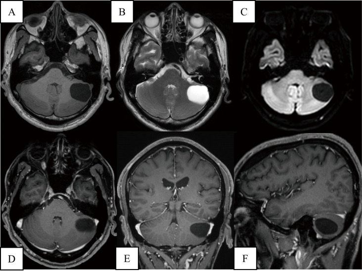
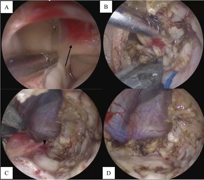
Intracranial cystic lesions such as hemangioblastoma (HB) are commonly found incidentally; however, they can be difficult to diagnose because they require various differential diagnoses. A contrast-enhanced mural nodule on magnetic resonance imaging (MRI) is typical and can be diagnosed preoperatively; however, some small nodules cannot be visualised and only cysts may be seen, complicating preoperative diagnosis. In such cases, thorough observation of the cysts is necessary for a definitive diagnosis. To achieve this, minimally invasive surgery, such as endoscopic keyhole surgery, is required. Herein, we report the case of a man in his 50s who presented with an unstable gait, and experienced dizziness for several months. Preoperative MRI revealed a cystic lesion in the left cerebellar hemisphere, without a mural nodule. Although there was no diagnostic evidence of HB, we suspected that the symptoms were caused by this cystic lesion because of its recent occurrence. Upon detecting a mural nodule, we diagnosed it as a cerebellar HB and completely resected it using an endoscopic keyhole approach. The patient's symptoms alleviated postoperatively. The endoscopic keyhole approach may be useful as a less invasive procedure for diagnosing and removing cystic cerebellar HBs, especially for lesions that are difficult to diagnose using preoperative imaging.


 分享
分享

 求助内容:
求助内容: 应助结果提醒方式:
 扫码关注我们
