Leonardo Bradaschia, Filippo Lacatena, Francesca Vincitorio, Paolo Titolo, Bruno Battiston, Diego Garbossa, Fabio Cofano
{"title":"Management of Post-Traumatic Pseudomeningocele as Consequence of Root Nerve Avulsion: Case Report and Review of the Literature.","authors":"Leonardo Bradaschia, Filippo Lacatena, Francesca Vincitorio, Paolo Titolo, Bruno Battiston, Diego Garbossa, Fabio Cofano","doi":"10.3390/neurolint16060126","DOIUrl":null,"url":null,"abstract":"<p><strong>Background: </strong>Post-traumatic pseudomeningoceles are common findings after a brachial or lumbar plexus trauma, in particular after nerve root avulsion. Unlike meningoceles, pseudomeningoceles are CSF full-filled cysts confined by the paraspinous soft tissue, along the normal nerve course, in communication with the spinal subarachnoid spaces. Normally no more than a radiological finding at MRI, in rare instances they might be symptomatic due to their size or might constitute an obstacle during a reconstructive surgery.</p><p><strong>Methods: </strong>A review of the literature was performed in accordance with the Preferred Reporting Items for Systematic Reviews and Meta-Analyses (PRISMA) guidelines in a time span ranging from November 1972 to May 2024. A total of five articles were found meeting the inclusion criteria. A case report at our institution was added to the case history.</p><p><strong>Results: </strong>A 30-year-old man with complete right brachial plexus nerve roots avulsion and a voluminous pseudomeningocele at the C6-C7 level after a motorcycle incident in January 2023. The pseudomeningocele covered the entirety of the injured brachial plexus. Pre-operative external lumbar drainage was utilized to prevent relapse or worsening of the already existing cerebral spinal fluid collection, with good results at 6 months. The full case report is reported in detail.</p><p><strong>Conclusions: </strong>To date, no clear guidelines about the management of post-traumatic pseudomeningoceles are reported in the literature. The lack of symptoms or signs related to them does not usually require any surgical intervention. If not, a possible management strategy with the use of an external lumbar drainage is proposed, a solution already in use in other surgical contexts with successful results in preventing CSF fistula or its relapse.</p>","PeriodicalId":19130,"journal":{"name":"Neurology International","volume":"16 6","pages":"1742-1749"},"PeriodicalIF":3.0000,"publicationDate":"2024-12-06","publicationTypes":"Journal Article","fieldsOfStudy":null,"isOpenAccess":false,"openAccessPdf":"https://www.ncbi.nlm.nih.gov/pmc/articles/PMC11679861/pdf/","citationCount":"0","resultStr":null,"platform":"Semanticscholar","paperid":null,"PeriodicalName":"Neurology International","FirstCategoryId":"1085","ListUrlMain":"https://doi.org/10.3390/neurolint16060126","RegionNum":0,"RegionCategory":null,"ArticlePicture":[],"TitleCN":null,"AbstractTextCN":null,"PMCID":null,"EPubDate":"","PubModel":"","JCR":"Q2","JCRName":"CLINICAL NEUROLOGY","Score":null,"Total":0}
引用次数: 0
Abstract
Background: Post-traumatic pseudomeningoceles are common findings after a brachial or lumbar plexus trauma, in particular after nerve root avulsion. Unlike meningoceles, pseudomeningoceles are CSF full-filled cysts confined by the paraspinous soft tissue, along the normal nerve course, in communication with the spinal subarachnoid spaces. Normally no more than a radiological finding at MRI, in rare instances they might be symptomatic due to their size or might constitute an obstacle during a reconstructive surgery.
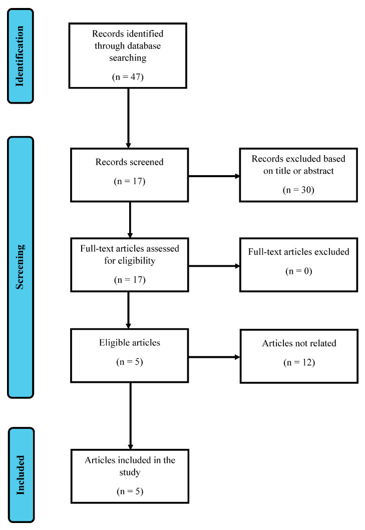
Methods: A review of the literature was performed in accordance with the Preferred Reporting Items for Systematic Reviews and Meta-Analyses (PRISMA) guidelines in a time span ranging from November 1972 to May 2024. A total of five articles were found meeting the inclusion criteria. A case report at our institution was added to the case history.
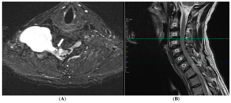
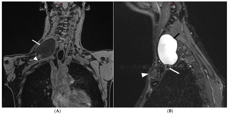
Results: A 30-year-old man with complete right brachial plexus nerve roots avulsion and a voluminous pseudomeningocele at the C6-C7 level after a motorcycle incident in January 2023. The pseudomeningocele covered the entirety of the injured brachial plexus. Pre-operative external lumbar drainage was utilized to prevent relapse or worsening of the already existing cerebral spinal fluid collection, with good results at 6 months. The full case report is reported in detail.
Conclusions: To date, no clear guidelines about the management of post-traumatic pseudomeningoceles are reported in the literature. The lack of symptoms or signs related to them does not usually require any surgical intervention. If not, a possible management strategy with the use of an external lumbar drainage is proposed, a solution already in use in other surgical contexts with successful results in preventing CSF fistula or its relapse.


 分享
分享
 求助内容:
求助内容: 应助结果提醒方式:
 扫码关注我们
